Ewing's Sarcoma
Findings:
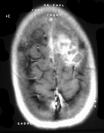
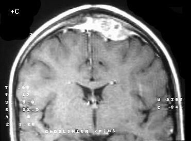
A plaquelike extraaxial soft tissue mass is present in
the left frontal calvarium which destroys the inner table. The mass has
irregular ill defined margins and enhances brightly with some heterogeneity.
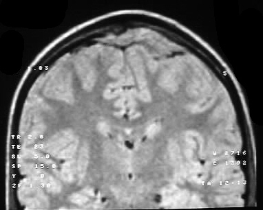
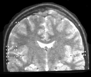
The mass has heterogenous isointensity on T2 weighted images, raising the
possibility of a cellular lesion. The T2 weighted images also show focal
disruption of the dura and a clear CSF space is not seen between the tumor
and brain parenchyma.
Differential Diagnosis:
At first glance, an en plaque meningioma may come to
mind. However, the characteristics described above would favor a more aggressive
lesion such as a sarcoma or metastatic disease. EG could have this appearance
as well. Due to the lack of brain atrophy, slitlike ventricles, and absence
of white matter disease, this is likely a young patient and Ewing's sarcoma
enters the differential.
Discussion:
Ewing's sarcoma may be seen in the age range of 1-30,
with 5-15 most common. Although a long bone origin is most common overall,
flat bones such as the skull and pelvis become more common as a site of
origin with increasing age. These tumors are small round blue cell tumors,
and may cause a permeative pattern with aggressive periostitis. An associated
soft tissue mass is usually seen, but this is not helpful to distinguish
between this and other processes such as EG and infection. The MR appearance
is nonspecific but helpful to determine extent of involvement and multifocality.