Germinoma
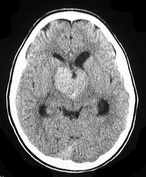
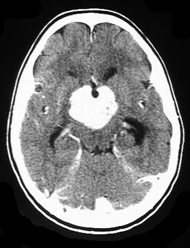
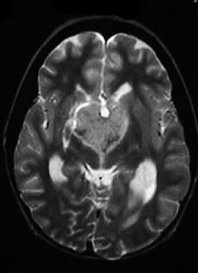
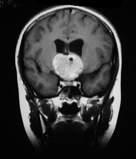
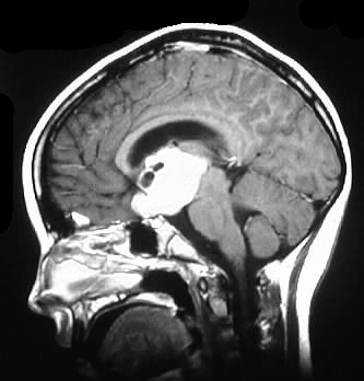
Findings:
A strongly enhancing solid suprasellar intraaxial mass
is present, which shows hyperdensity on precontrast CT adn heterogenous
slight hyperintensity on T2. Mild hypdrocephalus is evident as well. A
small cystic area is present in the periphery of this mass anterosuperiorly.
Differential Diagnosis:
Precontrast hyperdensity helps limit the differential
to germinoma and possibly lymphoma. Extraaxial masses such as meningioma
and craniopharyngioma could be considered as well.
Discussion:
Germinomas are in the family of germ cell neoplasms,
which also includes seminoma, embryonal carcinoma, yolk sac tumor, choriocarcinoma,
teratoma, and mixed tumors. Germinomas are most common in the pineal region
(65%), where there is a strong male predominance. 22% of germinomas are
found in the suprasellar region, and there is an equal male/female incidence
in this location. Peak incidence is around the age of puberty, suggesting
a hormonal influence. CSF dissemination and hydrocephalus are common, but
the tumors are highly radiosensitive and have a five year survival rate
in excess of 50%. Helpful imaging characteristics include precontrast hyperdensity
and occasional T2 hypointensity or mixed signal.
BACK TO
MAIN PAGE