Glomus Juguloympanicum
Findings:
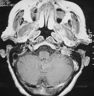
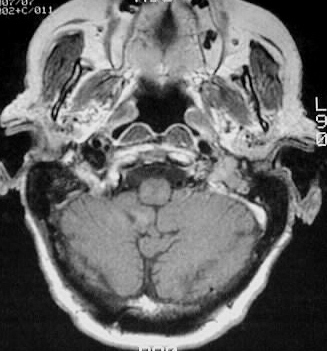
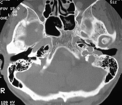
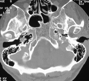
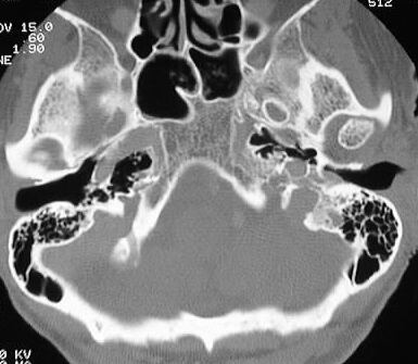
A soft tissue mass is present in the posterior portion
of the left middle ear, which abuts the carotid canal and extends into
the external auditory canal. The mass also involves the jugular foramen.
The MR images show strong enhancement of this mass, with stippled internal
hypointensities representing flow voids.
Differential Diagnosis:
glomus jugulotympanicum, cholesteatoma (atypical location),
squamous cell carcinoma. The MR appearance is fairly characteristic for
a glomus tumor.
Discussion:
Glomus tumors arise from rests of paraganglionic tissue,
but are infrequently biochemically active. Glomus jugulotympanicum commonly
originates in the area of the cochlear promontory, is more common in females
(2-4:1), and is the most common middle ear tumor. Clinically, these tumors
present as a vascular retrotympnic mass, and may cause pulsatile tinnitus.
Although the histology is benign, recurrence is common. 10% are multiple.
Hypervascularity accounts for the characteristic "salt and pepper" MR appearance,
with supply from the tympanic branch of the ascending pharyngeal artery
as well as other ECA branches. Irregular bone destruction is a characteristic
feature.