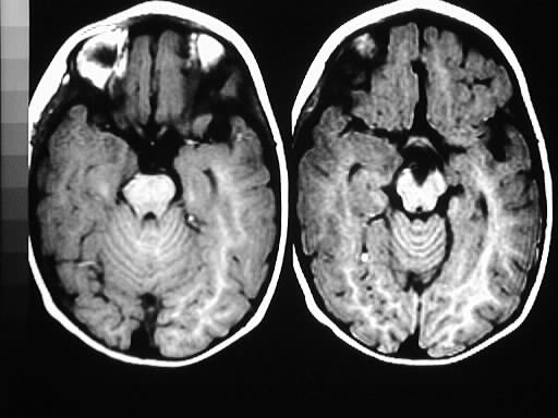
Findings:
Axial T1 images show an area of subtle hypointensity
in the right medial temporal lobe, without significant associated mass
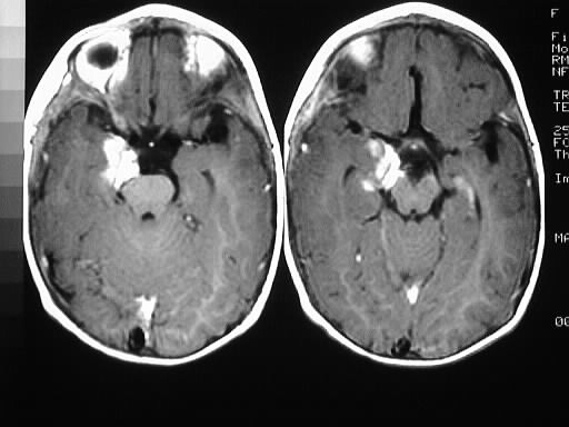
effect or edema. This area has bright gyriform enhancement.
Differential Diagnosis:
ganglioglioma, DNET, gangliocytoma, oligodendroglioma,
focal encephalitis or aging infarct less likely.
Discussion:
Gangliogliomas are low grade tumors composed of
glial cells and differentiated neurons, are most common in the temporal
lobes, and represent approximately 1% of intracranial neoplasms. Clinically,
these tumors present with partial complex seizures and/or hypothalamic
dysfunction at age 10-20. 5% are associated with additional anomalies (callosal
agenesis, Down's). The tumors are calcified in 35%, show no significant
mass effect or edema, and have variable enhancement.