

Neurofibromatosis type I
Findings:
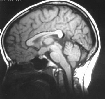
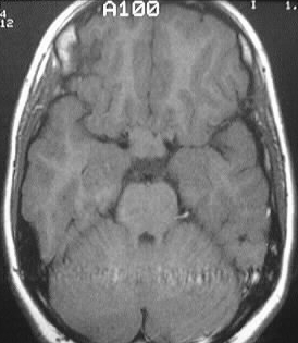
The optic chiasm is enlarged and lobulated, without significant
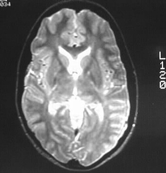
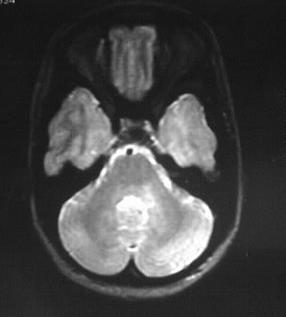
enhancement, consistent with optic glioma. Scattered hyperintense lesions
are present in the basal ganglia and left brachium pontis.
Differential Diagnosis:
Optic gliomas may arise sporadically in the absence of
NF I, but the associated parenchymal bright spots ensure a diagnosis of
NF I.
Discussion:
Neurofibromatosis type I- AD, 1/2500, 50% spontaneous
mutation
-clinical dx: 2 or more
-cafe au lait
spots (6 or more, >5mm child, >15 mm adult),
-NFs 2 or
more
-plexiform
NF
-axillary/intertriginous
freckles
-optic glioma
-lisch nodules
-bone lesions
-relative
with NF
-up to 50% of optic nerve gliomas assd with NF1
-cutaneous lesions
-cafe au lait
spots (coast of California, McCune Albright coast of Maine)
-freckling
intertriginous
-NFs, (TNTC
NFs=fibroma molluscum)
-elephantiasis
neuromatosa
-bone findings
-macrocephaly,
lambdoid defect, enlarged neural foramina (with or without NFs)
-sphenoid
dysplasia
-posterior
vertebral scalloping and scoliosis
-pseudarthrosis
-genu valgum/varum
-ribbon ribs
-tumors
-optic nerve
glioma
-cord astrocytomas
-malignant
peripheral nerve sheath tumors
-embryonal
tumors, leukemia, melanoma, medullary thyroid cancer
-other CNS lesions
-bright spots
in GP- tend to regress
-cerebellar
hamartomas
-arachnoid
cyst, meningocele
-vascular lesions
-renal artery
stenosis
-smoothly
tapered stenosis/occlusion of visceral arteries
-coarctation
-intracranial
stenosis