Rathke's cleft cyst
Findings:
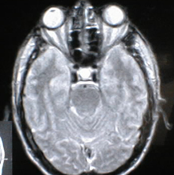
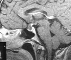
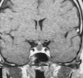
A round fluid signal mass is present in the sella, extending
into the infundibular region. The postcontrast images show minimal enhancement
around the rim of the mass.
Differential Diagnosis:
Rathke's cleft cyst, craniopharyngioma, cystic pituitary
adenoma less likely
Discussion:
Craniopharyngioma and Rathke's cleft cyst both arise
from squamous epithelial remnants of anterior lobe. Craniopharyngioma arises
from the pars tuberalis and Rathke's arises from pars intermedia. Rathke's
cleft cysts are 2/3 intrasellar and 1/3 suprasellar, contain fluid which
is mucoid, serous, or has cellular debris, and have variable MR signal
depending on composition. 50% may have minimal peripheral enhancement,
but the degree of enhancement is usually on of the most helpful factors
to distinguish this lesion from craniopharyngioma.
BACK TO
MAIN PAGE