Tectal Glioma
Findings:
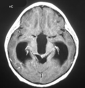
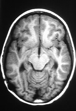
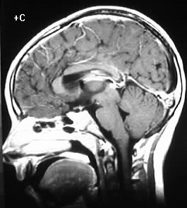
The lateral and third ventricles are markedly dilated
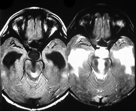
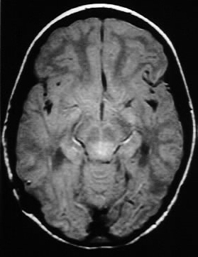
and the fourth ventricle is normal in size. The FLAIR and T2 weighted images
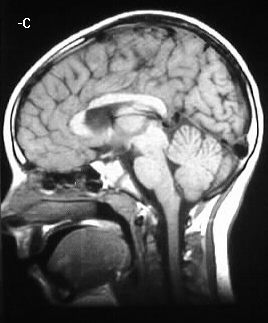
show a focus of abnormal signal in the region of the cerebral aqueduct.
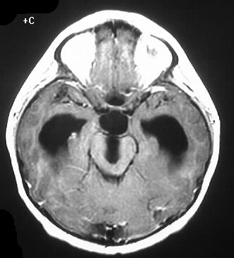
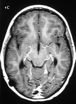
There is no definite abnormal enhancement. The follow up images show a normal ventricular system after shunting, with unchanged dorsal midbrain signal abnormality.
Differential Diagnosis:
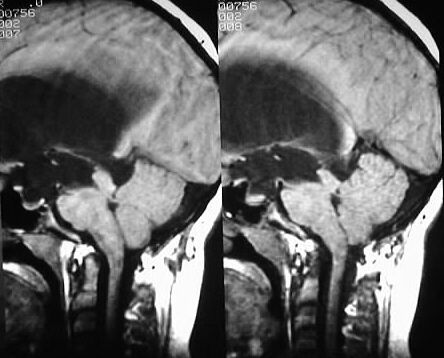
An obstructing lesion is present at the level of the
cerebral aqueduct which causes noncommunicating hydrocephalus. Differential
diagnostic possibilities include a small glioma and aqueductal stenosis
in a child. In an adult, etiologies such as glioma, metastasis, and possibly
lymphoma should be considered.
Discussion:
Tectal gliomas usually have low grade histology, but
are not amenable to resection due to the strategic location. In many cases,
shunting is all that is needed to allow long term survival.