Vein of Galen Malformation
Findings:
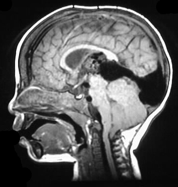
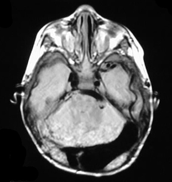
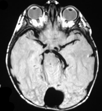
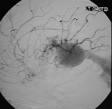
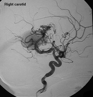
MR images show a massively enlarged Vein of Galen, stright
sinus, and torcula, associated with markedly enlarged posterior cerebral
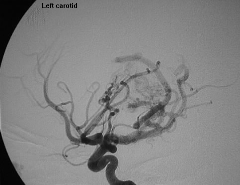
arteries. The arteriogram shows a nidus of vessels in the region
of the posterior third ventricle, with a large aneurysmal draining venous
structure.
Discussion:
Classification:
I- direct AV fistula between arteries
and VoG
II- thalamic AVM
III- mixture of I and II- neonate
with CHF
IV- AVM with veins that drain into
VoG
70% have associated venous anomalies (absent/interrupted
straight sinus, persistent falcine sinus). Posterior choroidal, ACA, PCA,
thalamoperforators, perimesencephalic vessels are most common arterial
supply. Associated syndromes include Turner's and Gorlin's (BRBNS).
reference: Osborn, A., Tong, K. Handbook of Neuroradiology: Brain and Skull. 1996:Mosby-Year Book, pp. 365-66.