Dural AV Fistula
Findings:
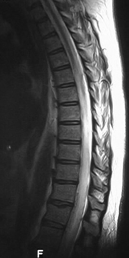
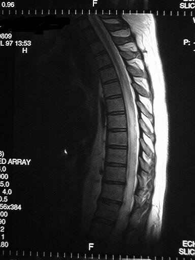
Sagittal T2 weighted images of the thoracolumbar spine
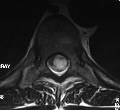
show diffuse abnormal hyperintense intramedullary signal, associated with
a few serpentine low voids along the dorsal surface of the spinal cord.
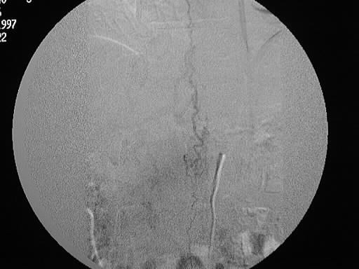
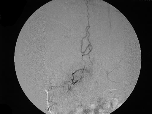
Spinal angiography shows abnormally enlarged draining veins at the level
of L1, associated with a small nidus of vessels.
Differential Diagnosis:
The differential diagnosis of abnormal intramedullary
signal includes MS, transverse myelitis, tumor, trauma, ischemia/infarction
and venous hypertension due to vascular malformation. The extent of this
abnormal signal would make MS and trauma unlikely. The presence of this
diffuse abnormal signal should raise the possibility of underlying vascular
malformation, especially if there is a history of longstanding progressive
myelopathy. Tumor must also be considered.
Discussion:
Classification of spinal vascular malformations:
I- nidus adjacent to or within dura
in region of proximal nerve root- most common
-Foix-Alajouanine syndrome- venous hypertension with progressive myelopathy
II- intramedullary- acute symptoms
due to hemorrhage
III- juvenile- extensive intra/extramedullary-
poor prognosis
IV- intradural extramedullary
Dural AVMs usually present with progressive myelopathy
in males 50-60s and may be difficult to detect if subtle flow voids are
not appreciated.