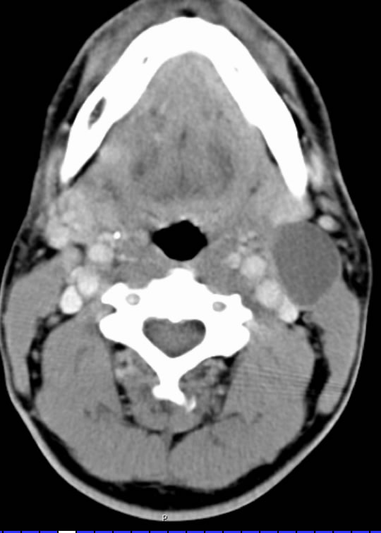
2nd Branchial Cleft Cyst
Findings:
Axial postcontrast CT neck shows a well defined cystic lesion in the left jugulodigastric region with very minimal uniform rim enhancement and internal slight hyperdensity to simple water.
Differential diagnosis:
branchial cleft cyst, nodal metastasis, suppurative adenopathy less likely
Discussion:
-2nd is most common- situated between SM gland and SCM at mandibular angle level
-1st is located in the parotid gland anterior to the external ear
-3rd is located in posterior cervical space
-Round unilocular simple fluid attenuation with very thin uniform wall
-More complex appearance if infected- also consider necrotic node(s) in these cases
-Don’t bet against squamous cell carcinoma if over 40
BACK TO
MAIN PAGE