Brain Abscess
Findings:
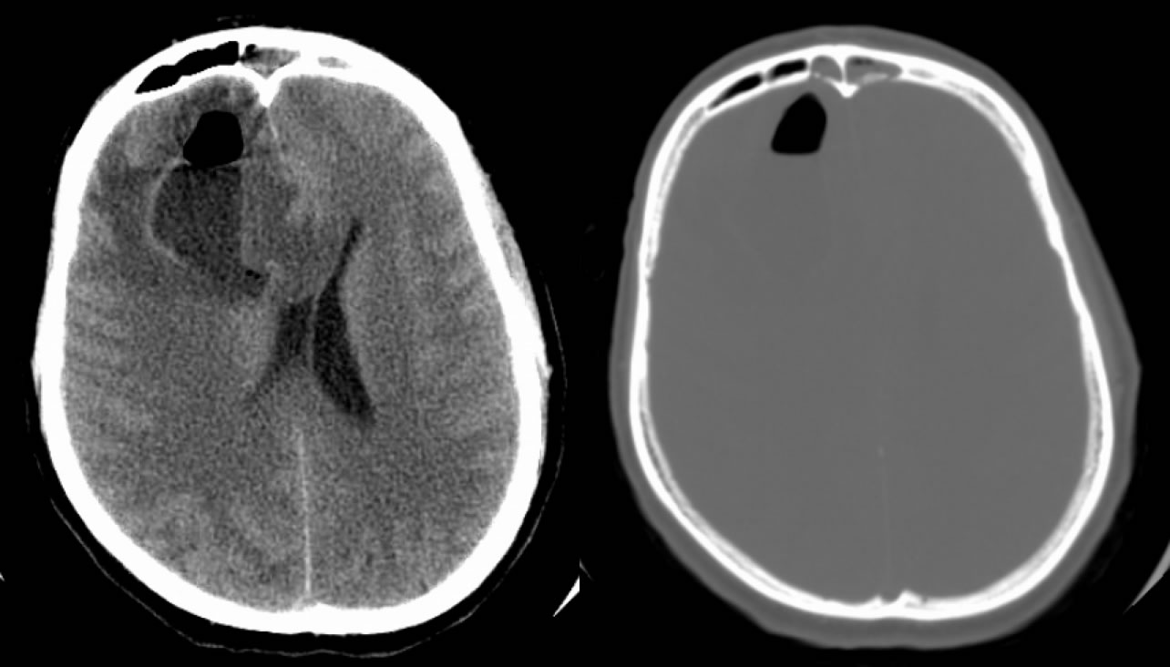
Axial noncontrast head CT demonstrates an abnormal fluid collection in the right frontal lobe with an air fluid level and surrounding vasogenic edema, associated with moderate mass effect and ventricular effacement. Bone windows show erosion of the frontal sinus inner table with inflammatory opacification of the frontal sinuses.
Differential diagnosis:
Brain abscess, necrotic neoplasm (AFL not seen), trauma with contusion and pneumocephalus (unlikely).
Discussion:
-early cerebritis (3-5d), late cerebritis (2 wks), early capsule (>2 wks), late capsule (wks-mos)
-only 50% have fever or WBC count, 90% have headache, most have high ESR
-less than 2.5 cm may be treated with Abx alone, greater need drainage
-internal DWI hyperintensity characteristic, some help in dist GBM/mets from abscess, not absolute
-DWI may be helpful in f/u, development of hyper DWI may indicate recurrence
-T2 hypointense rim with late cerebritis and early capsule, also not absolute
BACK TO
MAIN PAGE