Cysticercosis, racemose form
Findings:
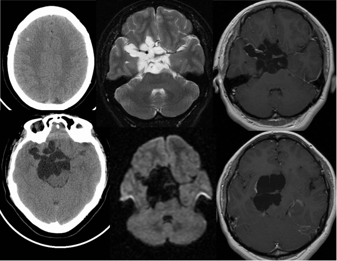
Multiple CT and MR images demonstrate multilobulated and multiseptated CSF signal/density structures within the suprasellar cistern, associated with localized mass effect and a few liniear areas of peripheral enhancement. A small hyperdense lesion is also visible in the right fromtal lobe. There is no restricted diffusion.
Differential diagnosis:
Racemose cysticercosis, epidermoid is primary differential (less likely with no diffusion restriction), complex arachnoid cyst less likely.
Discussion:
Racemose form is less common than parenchymal cysts. No scolices are seen. Calcification is not present and enhancement is minimal. Complications include hydrocephalus, meningitis, and ventriculitis.
BACK TO
MAIN PAGE