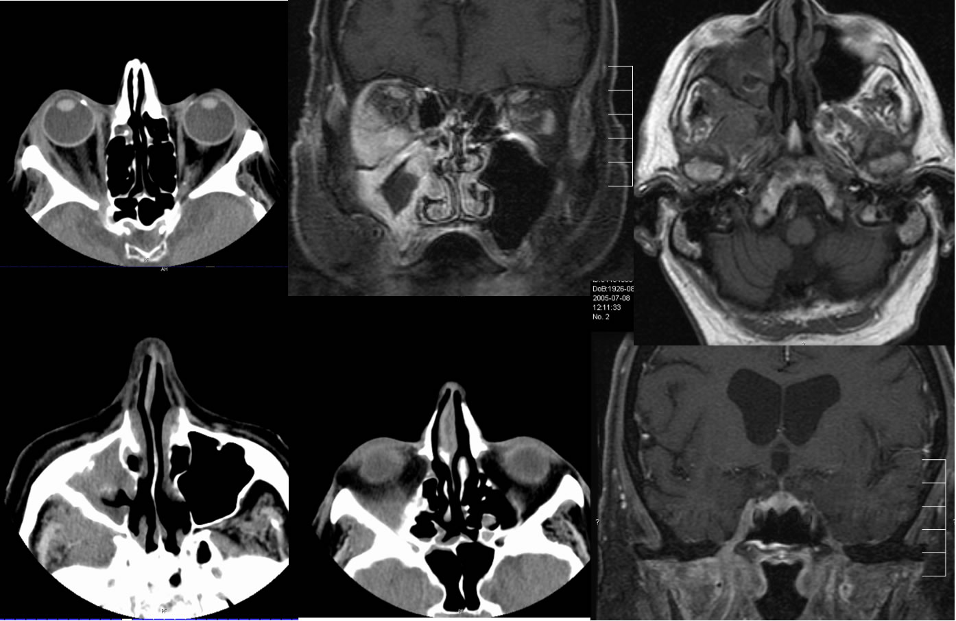
Invasive fungal sinusitis
Findings:
Multiple CT and MR images demonstrate complex opacification and signal abnormalities associated with the right maxillary sinus, with periantral fat plane and orbital extraconal postseptal infiltration. Abnormal enhancement also extends through foramen ovale. The right maxillary sinus walls are thickened but not destroyed.
Differential diagnosis:
Invasive fungal sinusitis, aggressive bacterial sinusitis, infiltrative neoplasm with perineural extent.
Discussion of fungal sinusitis:
nRange from nonspecific mucosal thickening to aggressive masslike appearance- Aspergillus most common organism
nLook for bone changes(sclerosis, erosion), hyperdense thick material, intrasinus calcifications, nasal mucosal thickening
nPeriantral soft tissue infiltration specific, but insensitive
nFungal hyphal colonization- range from near air density to dense fungus ball, early colonization may be asymptomatic
nAir fluid levels uncommon
nFour types: acute invasive (fulminant), chronic invasive, noninvasive colonization, allergic
BACK TO
MAIN PAGE