Frontal Sinus Mucocele
Findings:
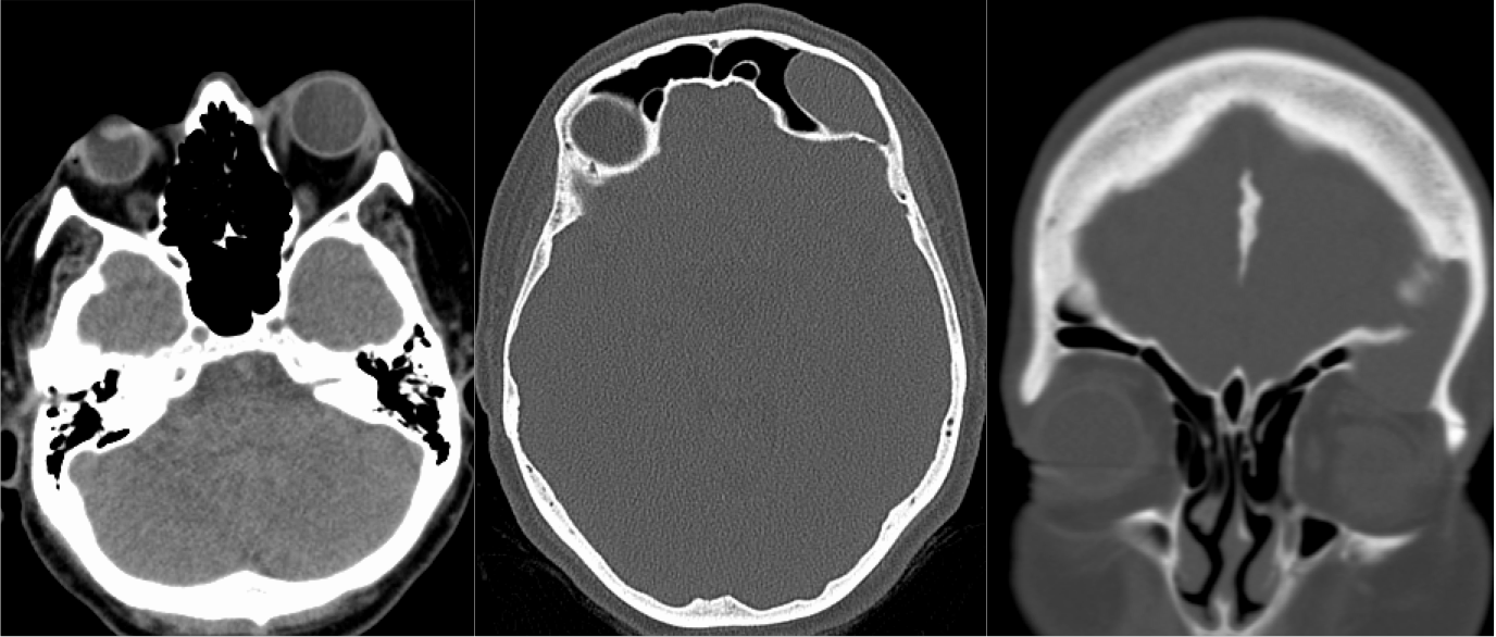
Noncontrast CT demonstrates an expansile lesion of the left fromtal sinus which causes proptosis and is associated with a well defined defect in the orbital roof. A thin margin of remodeled frontal sinus septum indicates a long standing process. The internal attenuation is in the region of complex fluid.
Differential diagnosis:
Appearance is fairly characteristic for mucocele. Low grade indolent neoplasm may appear similar.
Discussion:
Mucocele is one of three possible outcomes of chronic ostial obstruction. The other two include inspissation of secretions with atelectasis of the sinus and sinus wall thickening, and reestablishment of patency when inflammation reduces. Contents of a mucocele may have variable density and signal depending on proportion of mucinous goblet cell output versus serous fluid and balance of reabsorptive processes. Bone destruction is due to pressure erosion and takes some time, therefore there is bony remodeling rather than aggressive destruction.
BACK TO
MAIN PAGE