Tumefactive Demyelination
Findings:
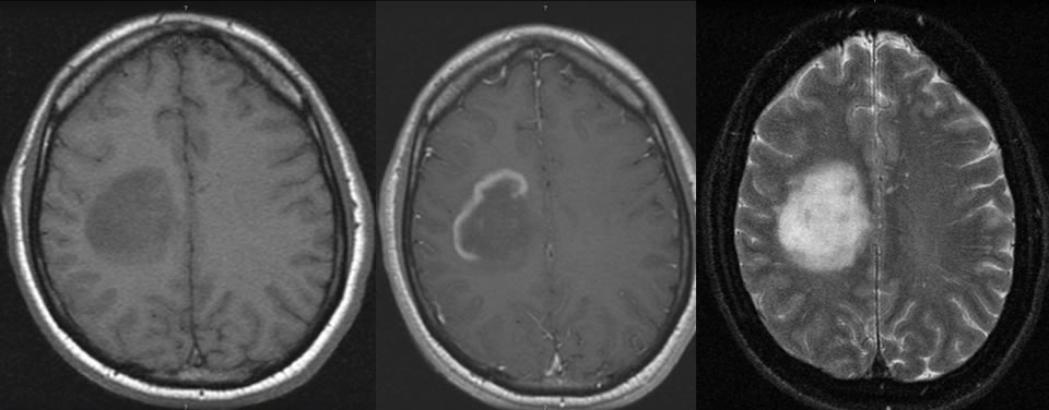
Axial T1 pre/post contrast and T2 fatsat images demonstrate an ovoid lesion in the right centrum semiovale that is not associated with mass effect. There is incomplete peripheral enhancement of the lesion. The associated T2 hyperintensity does not extend significantly beyond the leading edge of enhancement.
Differential diagnosis:
Demyelination, cerebritis (unusual without mass effect), primary or metastatic neoplasm (enhancement pattern is unusual for these, mass effect would be expected)
Discussion:
-Tumefactive type of MS may simulate neoplasm
-Look for incomplete ring enhancement
-Lack of edema/mass effect is characteristic
-Enhancing leading edge may be seen, with little signal change beyond leading edge
-Other white matter lesions may be seen that are more typical for MS
BACK TO
MAIN PAGE