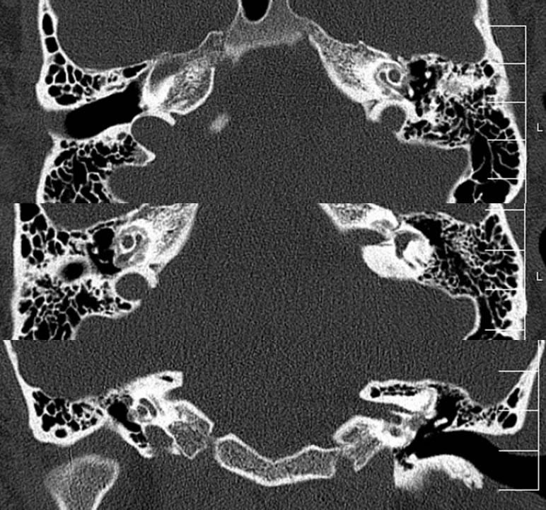
Otospongiosis/Otosclerosis, Retrofenestral
Findings:
Noncontrast temporal bone CT demonstrates a symmetric halo of demineralization surrounding the bilateral cochlea with subtle expansile appearance near the oval window niches, narrowing at least the left oval window.
Differential diagnosis:
Otospongiosis/otosclerosis, Osteogenesis imperfecta (more extensive demineralization expected)
Discussion:
-Many cases probably missed or not apparent on CT, particularly when fenestral and limited to anterior oval window margin
-Idiopathic vs. viral (measles), 85% bilateral, 2:1 F
-Initial demineralization, later proliferation/sclerosis
-Look for spongiotic plaque at anterior oval window
-Retrofenestral (cochlear) represents continuum with fenestral- cochlear rarely seen in isolation
-Obliterative form- thin plate of bone covering oval window (2%)
-Stapes footplate fixation- occult on CT
-Osteogenesis Imperfecta can appear similar or identical, but more extensive demineralization
BACK TO
MAIN PAGE