
Malignant (Necrotizing) Otitis Externa
Findings:
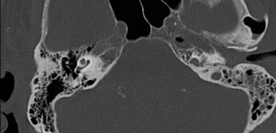
Noncontrast temporal bone CT demonstrates abnormal opacification of the left mastoid air cells and middle ear cavity as well as the external auditory canal. The soft tissue windows show infiltration of the left parapharyngeal and peristyloid fat planes.
Differential diagnosis:
Infection (MOE or uncomplicated otitis), trauma, infiltrative neoplasm (less likely)
Discussion:
-Invasive pseudomonal infection- junction of bony/cartilaginous EAC
-Elderly diabetics (93%)
-Rapidly progresses along undersurface of skull base, CN palsies VI-XII, sinus thrombosis
-Look for obliteration of fat planes beneath skull base- peristyloid, parapharyngeal, intermuscular
-Absence of bone destruction does not exclude MOE- obscured fat planes more sensitive
BACK TO
MAIN PAGE