Anaplastic Astrocytoma
Findings:
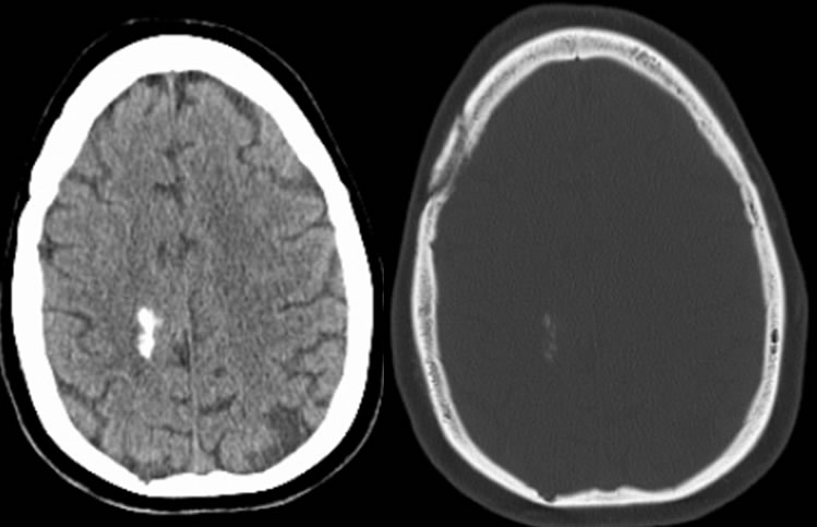
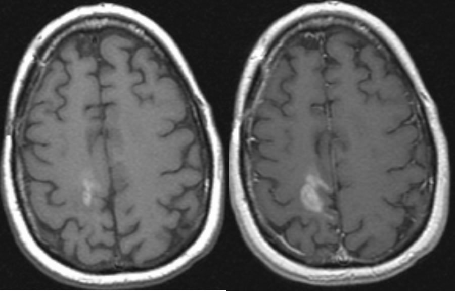
Axial noncontrast CT in brain/bone windows and T1 pre/postcontrast MR demonstrate a partially calcified lesion with associated nodular enhancement in the right frontoparietal parasagittal lobe. A remote craniotomy is incompletely included.
Differential diagnosis:
Oligodendroglioma, astrocytoma, metastasis.
Discussion:
Psammomatous calcifications may be seen in up to 25% astrocytomas, but are less common than calcifications in oligodendroglioma (90%), supratentorial PNET (60%), and ependymoma (50%)
nWHO classification of brain neoplasms 1993
grade I
-Juvenile pilocytic astrocytoma, pleomorphic xanthoastrocytoma, subeependymal giant cell astrocytoma, ganglioglioma
grade II
-diffuse astrocytoma, hemangiopericytoma
grade III
-anaplastic astrocytoma, hemangiopericytoma
grade IV
-glioblastoma multiforme (GBM)
BACK TO
MAIN PAGE