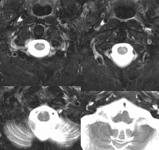
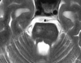
Multiple axial T2 weighted MR images demonstrate T2 hyperintensity and volume loss of the central right pons and olivary nucleus, with additional signal abnormality in the left lower cervicomedullary junction.
Differential Diagnosis:
Volume loss and T2 hyperintensity indicates a remote insult which may be due to many causes. If the anatomy of the corticospinal tract is not recognized, a misdiagnosis of MS or multiple separate remote infarcts might be made.
nVolume loss and gliosis distal to insult (treated brain tumor in this case)
nNeuroanatomy of corticospinal tract:
nPrimary motor cortex of precentralgyrusà coronaradiataà post limb ICàcerebralpedunclesà pontinetegmentum-à ventral medullary pyramidà decussate in caudal medulla-à lateral spinal cord, terminate in motor neurons of spinal cord gray matter