Right M1 segment occlusion with associated acute infarct
Findings:
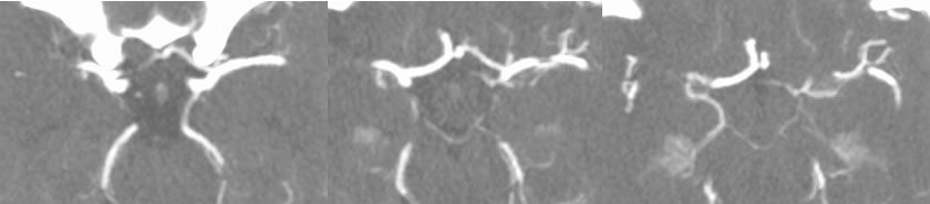
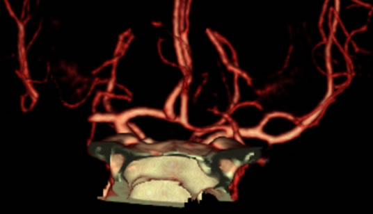
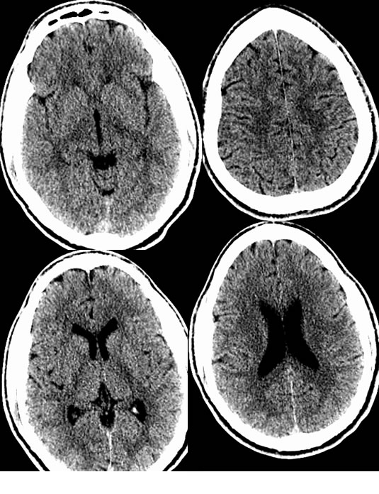
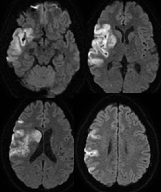
CTA demonstates abrupt occusion of the right M1 segment middle cerebral artery mid portion. CT demonstrates normal findings. DWI MR obtained 6 hours after the CT shows restricted diffusion in a large portion of the right insula, temporal lobe caudate, and frontoparietal opercular cortex along the right MCA distribution.
Discussion:
CT may be normal in the first several hours after vessel occlusion, while DWI will be positive within minutes. Subtle loss of grey white distinction of the external capsule and cortex may be the earliest visible CT sign of infarction. CTA is helpful for demonstration of the site of occlusion.
BACK TO
MAIN PAGE