Shear injury, Fornix
Findings:
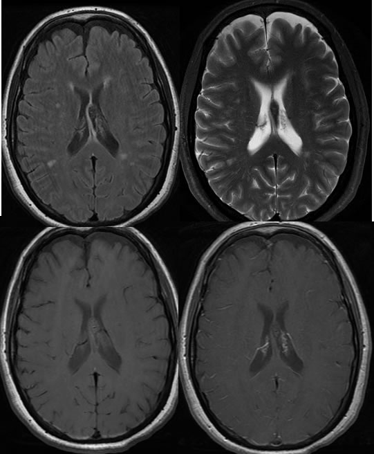
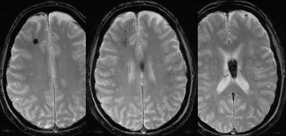
Multiple axial MR images demonstrate a T2 hypointense and T1 isointense lesion involving and slightly expanding the left forniceal column, without abnormal enhancement. GRE images demonstrate blooming of this lesion, as well as other spotty lesions with susceptibility in the right frontal white matter.
Discussion:
Diffuse axonal injury with or without hemorrhagic change is a common cause of morbidity in the setting of head trauma, and often results in long term disability. MR with T2* weighting is most sensitive for detection of shear injury. CT often underestimates the extent of DAI, but some shear injuries wil present as punctate hyperdensities in CT. Larger shear injuries, such as those involving the corpus callosum, will be visible on CT, as are many other manifestations of acute trauma.
BACK TO
MAIN PAGE