Tonsil Cancer
Findings:
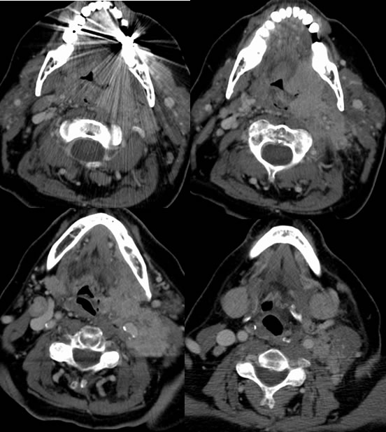
Multiple axial contrast enhanced CT images show a poorly defined infiltrative mass arising from the region of the left palatine tonsil, with extensive local invasion of the deep neck spaces and SCM. There are bilateral nodal metastases.
Differential Diagnosis:
None reasonable except malignancy, most likely squamous cell. High grade salivary gland malignancy might appear similar. The appearance is atypical for lymphoma, not excluded.
Discussion:
CT is important in the staging of head and neck malignancies. Tumor and nodal staging correlates with resectibility and prognosis. A tumor that invades local structures and deep neck spaces is considered T4. Bilateral adenopathy is considered N2c nodal metastatic disease. PET may be helpful to determine the extent of malignant adenopathy. T stages are at 2 cm intervals- T1 less than 2 cm, T2 2-4 cm, T3 greater than 4 cm. For nodes, any node greater than 6 cm makes N3, any node greater than 3 cm or more than one node is at least N2, and N1 is a single ipsilateral node less than 3 cm. N2a is a single node 3-6 cm, N2b is multiple ipsilateral nodes, and N2c is bilateral or contralateral nodes. Other details regarding AJCC TNM classification are readily available on Radiopaedia and Medscape.
BACK TO
MAIN PAGE