Epidermoid
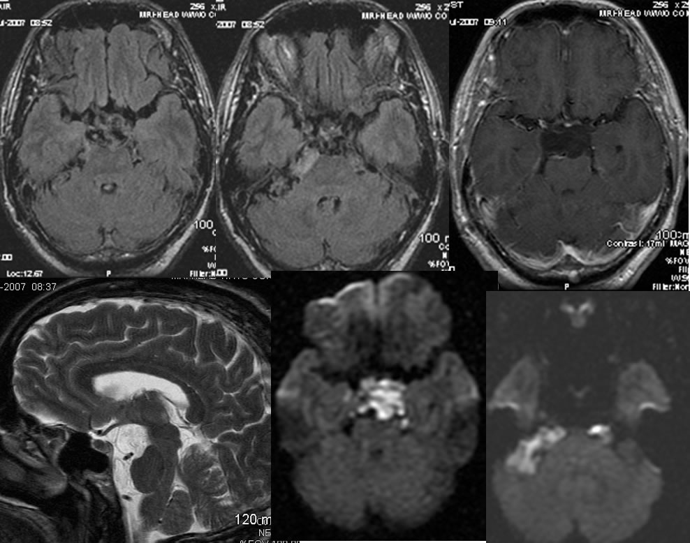
Findings:
Multiple MR images demonstrate irregular extraaxial masses in the suprasellar and prepontine/CP angle cisterns which show no abnormal enhancement and heterogenous FLAIR hyperintensity. Diffusion weighted imaging shows high signal in these regions. The lesion causes mild mass effect on the pons.
Differential Diagnosis:
Epidermoid, complex arachnoid cysts, racemose cysticercosis, sequelae of remote hemorrhage with septated extraaxial collections
Discussion:
nImaging
n90% intradural, 40-50% CPA, 17% IV vent, 10-15% parasellar, rare intraaxial (1.5%)
nCT
nCSF density, lobulated with mass effect, 15-20% calcification, <5% variable density due to hemorrhage, protein, saponification
nMR
nSlight hyper to CSF on T1 (variable), slight hyper on FLAIR, DWI hyper is key to dx., less common linear peripheral enhancement
nPath
nCongenital ectodermal inclusion, progressive internal desquamation
n“mother of pearl” grossly, filled with soft material
nFibrous capsule, epithelial lining, filled with cholesterol and keratin debris
nClinical
nHeadaches, CN neuropathy, DI, Sz, asymptomatic, peak 40 yrs.
nSlow growth, rare malignant degeneration to SCCa, chemical meningitis
nMicrosurgical resection- difficult due to insinuation with CN
BACK TO
MAIN PAGE