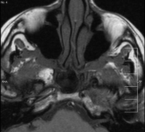
Perineural tumor spread, squamous cell carcinoma nasopharynx
Findings:
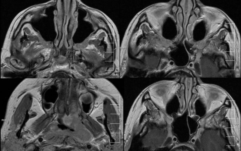
The initial image is axial T1 with no contrast or fatsat. This image shows symmetric infiltration of the bilateral pterygomaxillary fat planes. The additional images are axial T1 with contrast, which demonstrate the primary malignant neoplasm in the left nasopharynx, as well as the perineural tumor infiltration of the pterygomaxillary fissures and bilateral foramen rotundum.
Differential Diagnosis:
perineural tumor spread, extracranial spread of meningioma or other extraaxial mass, therapy related changes less likely.
Discussion:
nSquamous cell perineural tumor most common, Adenoid cystic “classic” but less common statistically
nOthers to show perineural spread: melanoma, lymphoma, mucoepidermoid, rarely mets
nComplete surgical resection is best chance of cure in H/N cancer- but more difficult or impossible if there is perineural spread
nSpread common along V3, V2, VII, rarely V1(skin, lacrimal)
nAreas to inspect closely in region of primary neoplasm
nMax sinus, nasopharynx à pterygopalatine fossa, foramen rotundum
nMasticator space, oral cavity à foramen ovale, inferior alveolar canal
nParotid à stylomastoid foramen, auriculotemporal nerve to V3 (interpterygoid fat plane, posterior mandible)
BACK TO
MAIN PAGE