Giant pseudoaneurysm, left internal maxillary artery
Findings:
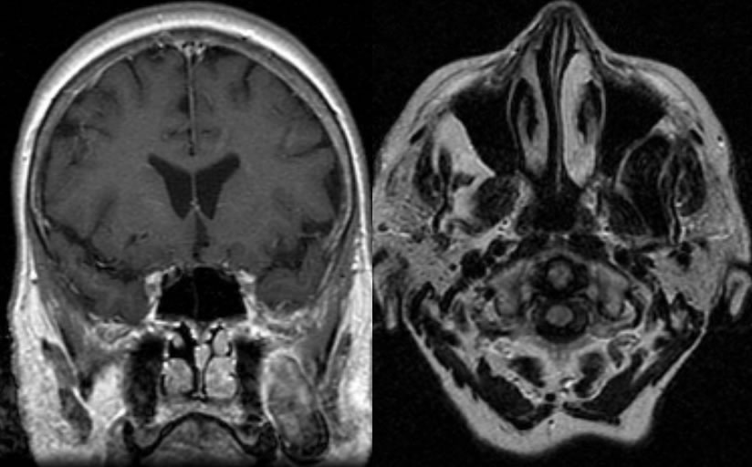
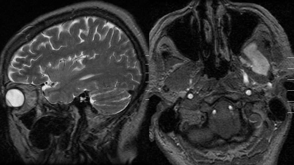
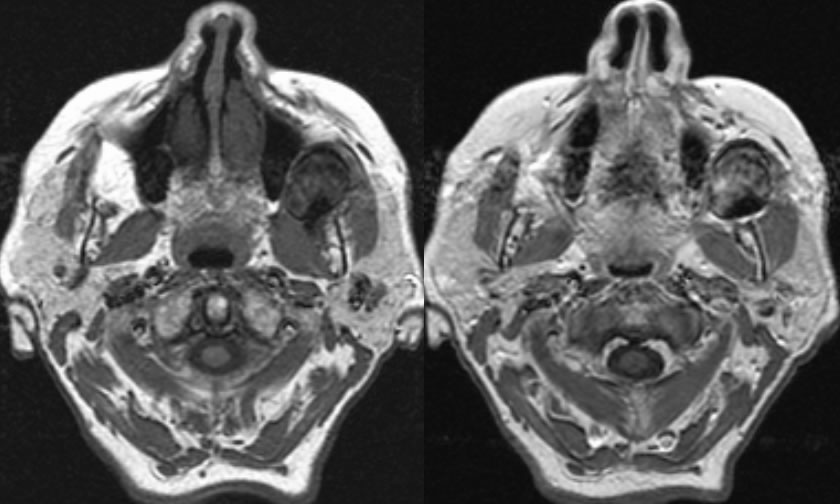
Multiple MR and MRA images demonstrate a lobulated left retromaxillary lesion with low signal on T2, internal arterial flow signal on MRA, and pulsation artifacts through the lesion on postcontrast T1 indicating a high flow vascular lesion.
Differential diagnosis:
Aneurysm, pseudoaneurysm, arteriovenous malformation, venous malformation
Discussion:
Spontaneous pseudoaneurysms are rare in the absence of trauma or regional surgery. This lesion was cured with embolization.
BACK TO
MAIN PAGE