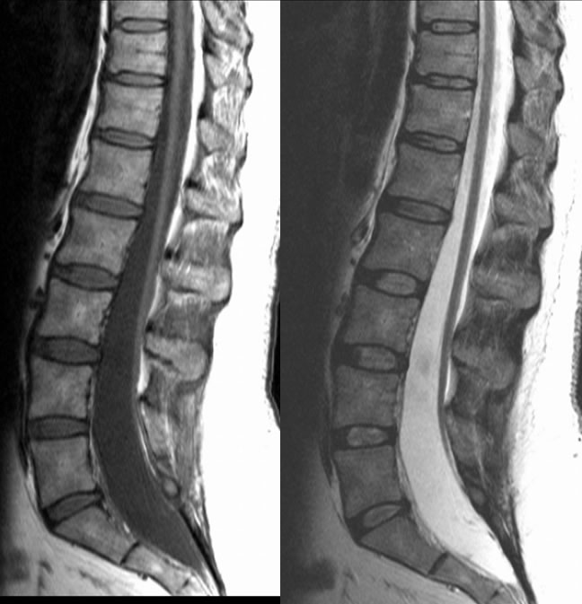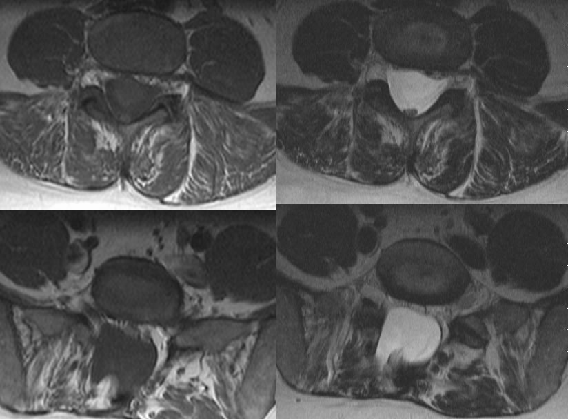

Lipomyelocele with tethered cord
Findings:
Multiple MR images show an elongated conus with tip not identified, likely near the S1 level. The spinal cord is diplaced and tethered into a fat signal mass associated with a low lumbosacral dysraphic defect.
Differential Diagnosis:
lipomyelocele, myelomeningocele, myelocele, postoperative changes.
Discussion:
nSpectrum of abnormalities from incomplete neural tube closure and failure of separation between neural and superficial ectodermal elements
nMost extreme: anencephaly
nIntermediate severity: Chiari II/myelomeningocele
nLeast severe: Dorsal dermal sinus
BACK TO
MAIN PAGE


