Schwannoma
Findings:
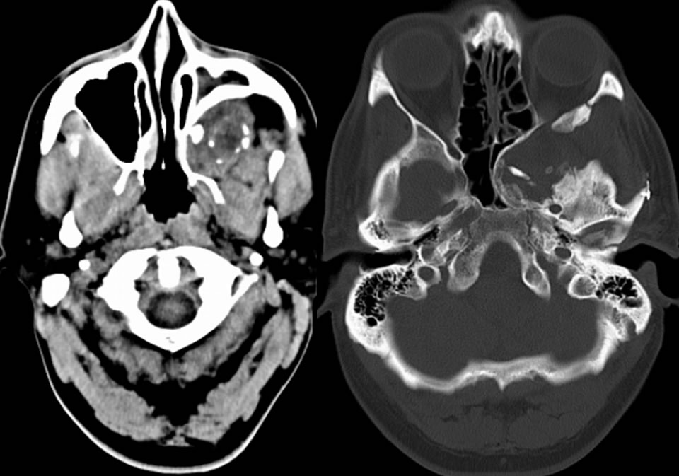
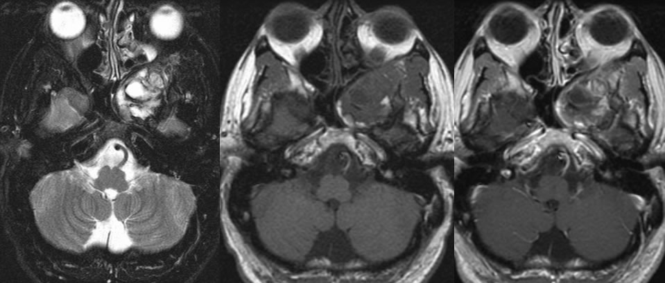
Multiple CT and MR images demonstrate a partially calcified left retromaxillary mass that is associated with extensive remodeling of the maxillary posterior wall indicating a long standign process. MR signal characteristics are heterogenous with complex cystic zones adn patchy nodular enhancement.
Differential Diagnosis
(Ancient) Schwannoma, thrombosed aneurysm, low grade salivary gland neoplasm
Discussion:
nClues that it is a neural origin tumor
nExtensive remodeling of skull base due to longstanding slowly growing tumor
nPredominantly T2 hyperintense with cystic areas and peripheral enhancement
nChunky and rimlike calcifications
nFollows/expands skull base foramina- rotundum
BACK TO
MAIN PAGE