Pontine capillary telangiectasia with DVA
Findings:
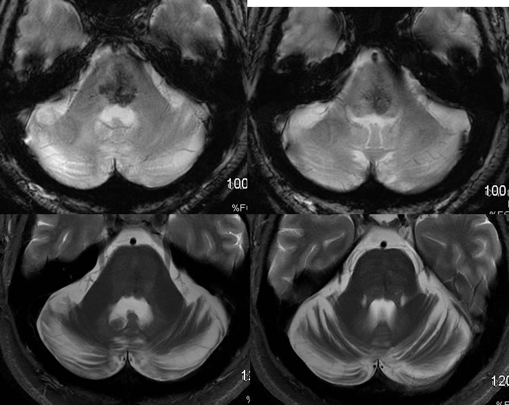
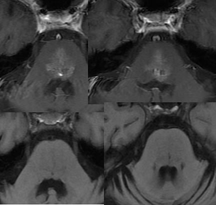
Multiple MR images show a large focus of patchy GRE hypointensity in the central pons, with no significant associated T2 hyperintensity. This structure demonstrates corresponding patchy enhancement and is essentially invisible prior to contrast administration. There is no definite expansion of the pons in this region.
Differential diagnosis:
Location and signal characteristics are most compatible with a benign incidental vascular anomaly such as telangiectasia/DVA. If there were more atypical features, neoplasm or brainstem hemorrhage could be considered.
Discussion:
nPons is most common location
nNot usually visible on T1 pre or T2/FLAIR
nFaint patchy enhancement
nGRE blooming-slow flow and desaturation
nNo mass effect or surrounding edema
BACK TO
MAIN PAGE