Dysembryoplastic Neuroepithelial Tumor (DNET)
Findings:
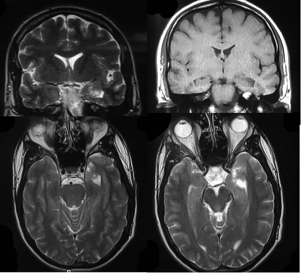
Coronal MR demonstrates a lesion in the medial left temporal lobe that is heterogenous and hyperintense on T2WI and hypointense on T1WI. Axial MR shows that the lesion does not exhibit mass effect, with a few spotty areas of bubbly T2 hyperintensity.
Differential Diagnosis:
DNET, Taylor dysplasia, hamartoma (TS), neuroepithelial cysts, ganglioglioma
Discussion:
Dysembryoplastic Neuroepitherlial Tumor (DNET) is a type of benign, focal, intracortical mass that is superimposed on a background of cortical dysplasia. Typical clinical history is that of intractable partial complex seizures in a child or young adult. There is a 100% association with complex partial seizures. These lesions have been reported in up to 80% of epilepsy specimens. Imaging with CT demonstrates a wedge-shaped area of low density that may resemble infarct, with no calcification. On MR they appear as a pseudocystic, multinodular mass which is hypointense on T1 and hyperintense on T2 with no significant enhancement, but some may havde a cyst and mural nodule appearance. Classic finding is that of a "bubbly" mass with septations best visualized on T2WI. A helpful imaging finding is an enlarged gyrus with normal white matter coursing into it. Hamartoma related to Tuberous Sclerosis can appear similar, but would be more commonly multiple with other findings of TS. The most common location for this type of lesion is in the temporal lobe, however the parietal cortex, caudate nucleus, and septum pellucidum would not be uncommon. DNET does not progress to malignancy and exhibits very slow, if any, growth. However, these lesions may need to be removed surgically if seizures persist. Pathologically, it demonstrates a polymorphic mixture of glioneuronal and cortical dysplastic features.
BACK TO
MAIN PAGE