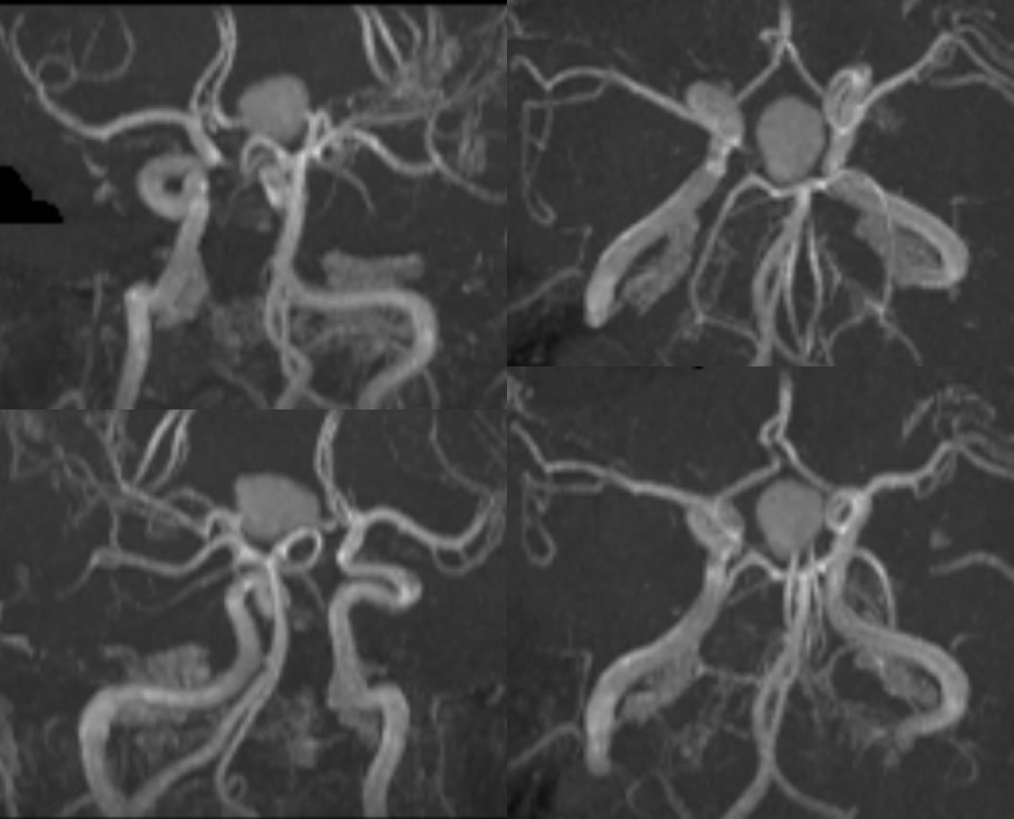
A rounded hyperintensity projects over the Circle of Willis on reformatted MRA images. The lesion has similar signal to surrounding arterial flow. However, upon further inspection the lesion is completely separate from the arterial system (best seen on the second image), therefore ruling out the possibility of a large aneurysm.
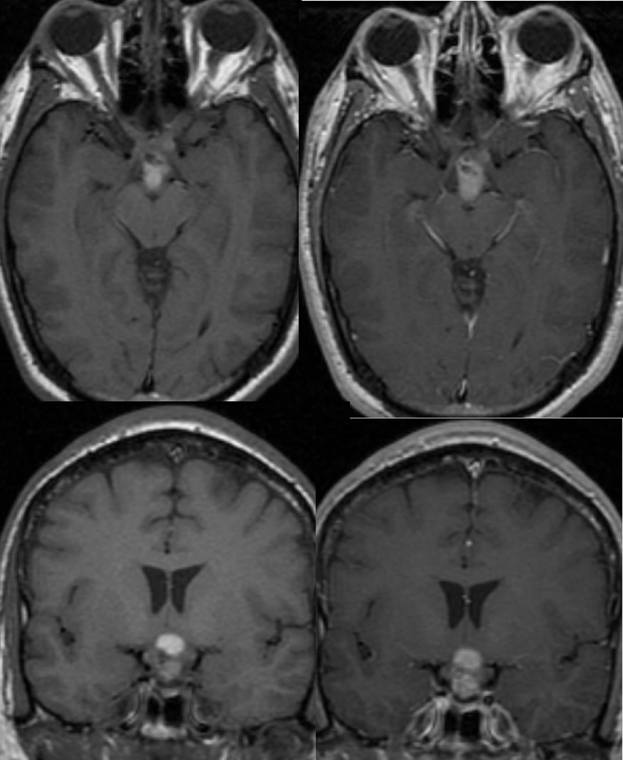
Axial and coronal pre/post contrast T1WI demonstrate a complex cystic mass in the suprasellar region. The mass is hyperintense, representing the cystic component, with some surrounding isointensity, representing the solid portion. Post-contrast images show that the lesion enhances in the area of the solid component with relative enhancement outlining the cystic portion.
Craniopharyngioma, complex Rathke cleft cyst, pituitary adenoma, other primary tumor in suprasellar region
Craniopharyngiomas are a type of benign epithelial tumor that arise from the remnants of Rathke's pouch. Clinical presentation is variable with the location and size of the mass, but may involve progressive headache, bitemporal hemianopsia from compression of the optic chiasm, and endocrine disturbances, particularly GH deficiency, from local extension into the sella turcica. On CT, craniopharyngiomas typically present as a partially calcified, partially solid, cystic suprasellar mass. MR shows a suprasellar mass that has a variable hyperintense cystic portion and a heterogenous nodular portion on T1WI. There is typically mild enhancement in the solid portion with strong cyst wall enhancement. Treatment is typically surgical and radiotherapy, but local recurrence is not uncommon. Overall all 10 year survival is good ranging from 64-96%.