
CNS Lymphoma
Findings:
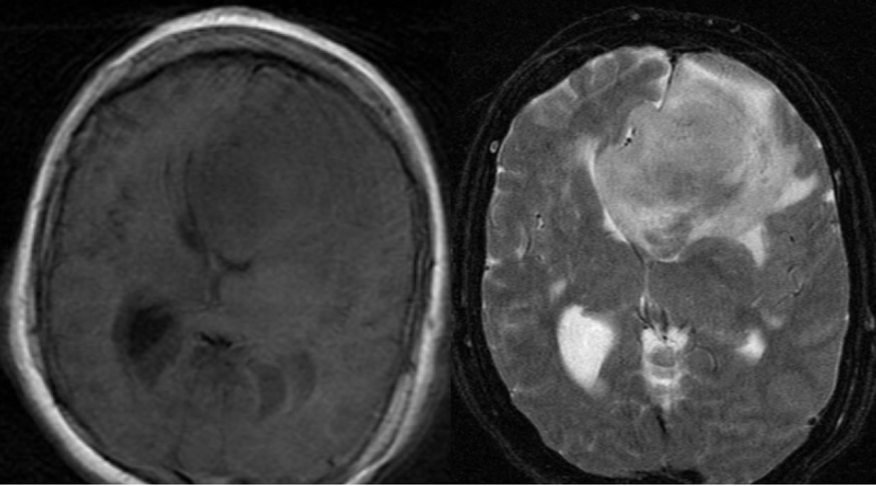
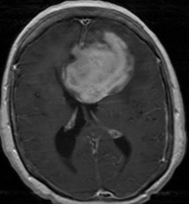
Axial T1WI demonstrates a hypointense mass in the left frontal region that demonstrates strong enhancement post-contrast. There is significant motion artifact in the T1 pre-contrast image. The mass is heterogenous and hyperintense on T2WI with surrounding vasogenic edema and causes rightward midline shift, compression of lateral ventricles, and sulcal effacement. Areas of relative low T2 signal are also present in the lesion indicating cellularity.
Differential Diagnosis:
Primary CNS lymphoma, metastatic tumor, glioblastoma multiforme, PML/IRIS
Discussion:
Primary CNS lymphoma is a malignant neoplasm composed of B lymphocytes. They account for 1-7% of primary brain tumors, and 1% of lymphomas. Ninety percent occur supratentorial, most commonly in the frontal and parietal lobes. These tumors commonly involve and cross the corpus callosum, and are known to abut and extend along ependymal surfaces. Classic imaging findings are that of a strongly enhancing lesion within the basal ganlgia or periventricular white matter. Classic CT finding is that of a hyperdense mass, although they may appear isodense. Imaging and prognosis varies with immune status. CNS lymphomas in immunocompetent individuals will show strong homogenous enhancement, while immunocompromised individuals tend to have a more aggressive form that will often show peripheral enhancement secondary to central necrosis. The average age at diagnosis is 60 years in immunocompetent and just under 40 years in the immunocompromised. Median survival ranges from 17-45 months among the immunocompetent down to 2-6 months in the immunocompromised.
BACK TO
MAIN PAGE


