Chondrosarcoma
Findings:
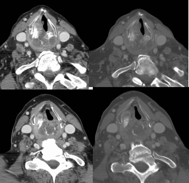
Axial contrast enhanced neck CT images in soft tissue and bone algorithm show an irregular expansile mass involving the cricoid and right arytenoid cartilages, which demonstrates predominantly low attenuation. Scattered curvilinear irregular calcifications are present, with thin peripheral enhancement.
Differential Diagnosis:
Chondrosarcoma, Chondroma, metastasis less likely.
Discussion:
nImaging
nExpansile mass arising from cricoid or laryngeal cartilage
nCT: calcified matrix (rings and arcs), otherwise hypodense to muscle, MR nonspecific hyper T2, T1 iso to muscle
nDDx
nChondroma, primary tracheal diseases (tracheopathia osteochondroplastica, relapsing polychondritis, laryngeal nodular chondrometaplasia), other sarcomas (not calcified)
nClinical/Path
nHyaline cartilage origin, not elastic, path grades I-III
n0.5% of laryngeal malignancies, mean 64, M>F 3.6:1
nAirway compromise
nMost low grade, but prognosis good despite grade (except for rare myxoid)
nSurgical resection of chondroid lesion usually curative whether benign or malignant
BACK TO
MAIN PAGE