Subacute Infarct
Findings:
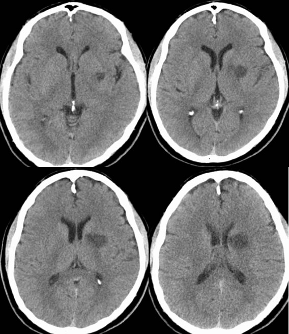
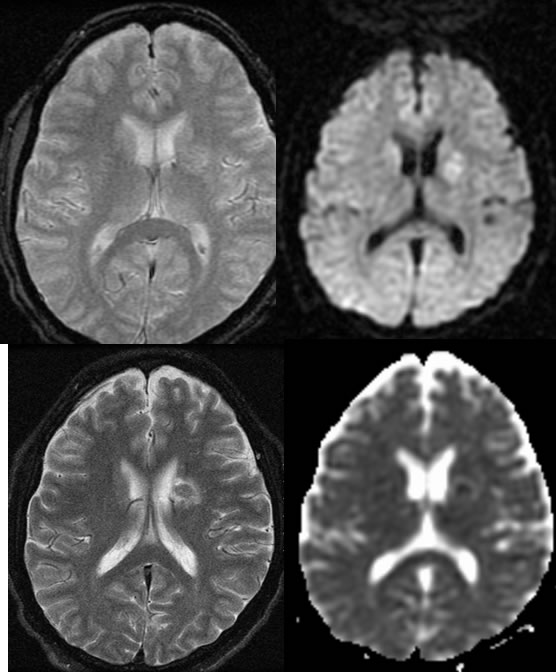
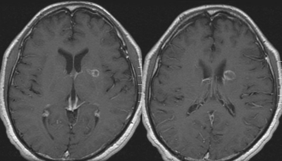
Axial noncontrast CT demonstrates a well defined wedge shaped focus of low attenuation involving the left corona radiata, caudate nucleus, and left basal ganglia. Multiple MR images show this lesion to have increased peripheral T2 signal with a central zone of T2 hypointensity. The lesion also show diffusion hyperintensity with decreased signal on ADC map. Gradient echo imaging shows no significant hemorrhagic staining. Mild peripheral enhancement is present.
nDDx
nDemyelination, glial or metastatic neoplasm, encephalitis
nImaging
nWedge shaped, mass effect diminishes after 7-10 days
nEnhancement patchy, peripheral, or gyriform begins 2-3 days and lasts up to 8-10 weeks
nKey feature
n2-2-2 rule- begins 2 days, peaks 2 weeks, resolves 2 months
nT2 fogging effect 6-14 days
nClinical
n25% mortality within 2 weeks due to recurrent stroke
46% 5ysr, 28% 10ysr (often from other comorbidities)
BACK TO
MAIN PAGE