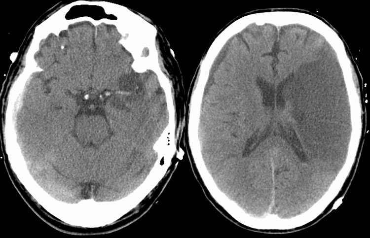
Left MCA Infarct with M1 segment thrombosis
nFindings
nThere is a large, well-established left MCA distribution infarct with a hyperdense M1 segment of the left MCA. In addition, there is a mixed density subdural hematoma over the right cerebral convexity.
nDifferential Diagnosis
nHyperdense Vessel: polycythemia/hemoconcentration, vessel wall microcalcification, consider thrombosis if asymmetric
nParenchymal Hypodensity: stroke, infiltrating neoplasm, aging cerebral contusion, inflammation
nDiscussion
nEvaluation of acute infarction by CT can be difficult due to lack of radiologic findings. In the hyperacute setting, there are typically no changes in brain attenuation. A specific, but only 35-50% sensitive, sign of acute infarction is a hyperdense vessel that signifies thrombosis. Other early signs include loss of gray-white differentiation within the first 3 hours and gyral swelling and sulcal effacement that become evident between 12-24 hours. Loss of the insular “ribbon” is a sign that signifies loss of gray-white matter border. As time progresses, parenchymal hypodensity will become apparent that correlates with the infarcted area. Hemorrhagic transformation occurs in 15-45% of cases with delayed onset (24-48 hours) being most common.
Case contributed by Christopher Heald, UC M4
BACK TO
MAIN PAGE