

Perched facets
nFindings
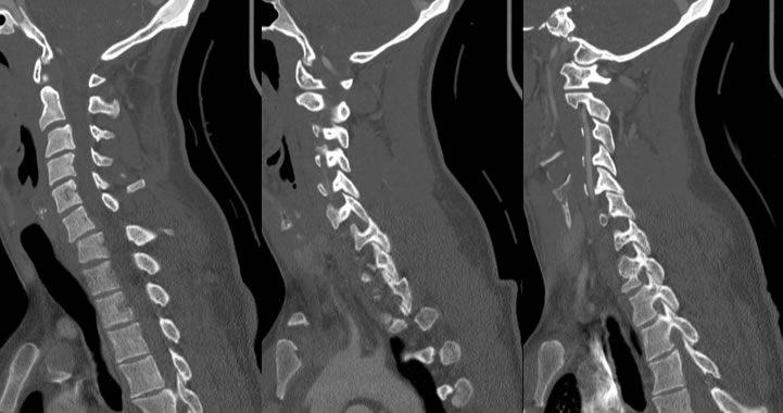
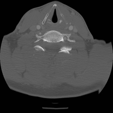
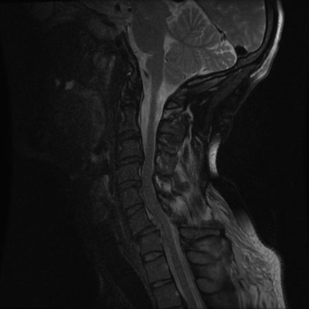
nSagittal CT demonstrates traumatic anterior subluxation of C6 on C7 with locked left and perched right facet joints. There are no acute fractures visible. There is increased soft tissue density along the posterior longitudinal ligament indicating possible disruption and uncovering of C6-7 disc. Axial CT demonstrates locked facet on the left, but perched facet on the right is difficult to visualize. Sagittal MR demonstrates discontinuity of the ligamentum flavum at C6-7. The posterior longitudinal ligament is not clearly disrupted, although it is displaced posteriorly. There is swelling in the anterior epidural space of C6-7 causing mild cord compression.
nDiscussion
nInjuries, such as those shown in the images above, occur secondary to traumatic hyperflexion of the cervical spine, classically during a motor vehicle accident. If trauma in this fashion is powerful enough, it may cause disruption of the ligamentous structures of the cervical spine allowing anterior subluxation of vertebral bodies and perched or locked facet joints. A perched facet describes a situation in which the inferior articular process is anteriorly subluxed and now resting on the anterior aspect of the superior articular process. Any further subluxation will cause dislocation of the joint, with the inferior articular process jumping over the superior articular process and becoming locked. In these situations there is concern for spinal cord compression, and this patient demonstrated mild compression from anterior swelling. The posterior longitudinal ligament is important in preventing hyperflexion injuries of the cervical spine and may become disrupted with a forceful hyperflexion. This ligament appears dark on T2WI and runs just posterior to the vertebral bodies.
Case contributed by Christopher Heald, UC M4
BACK TO
MAIN PAGE


