Otosclerosis with obliterative disease
Findings:
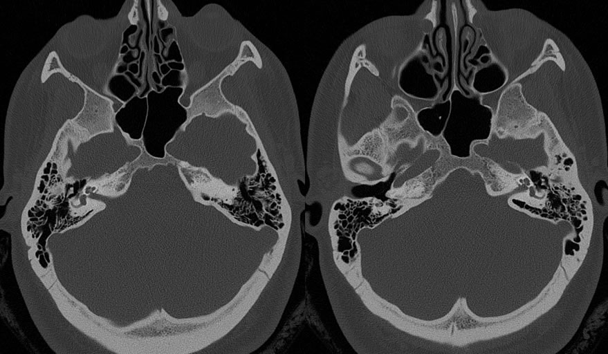
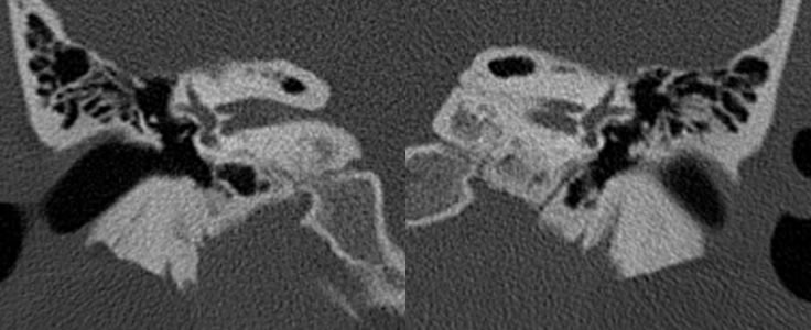
Axial CT demonstrates subtle lucencies along the bilateral oval window margins. There is also a halo lucency surrounding the left cochlea and an incomplete halo lucency surrounding the right cochlea. Coronal CT images show that there is markedly narrowed and subtotally obliterated right oval window and a partially obliterated left oval window.
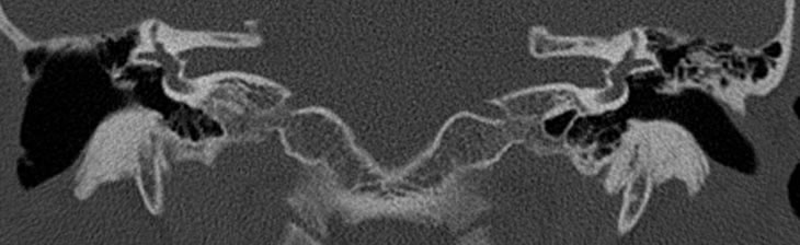
The third image was taken from a different patient who does not exhibit signs of otosclerosis. It has been added for comparison to demonstrate normal versus obliterated oval windows. Note that the comparison patient has had a prior right mastoidecomy.
Discussion:
Otosclerosis is an idiopathic disease of the perifenestral bony labyrinth in which spongy bone foci appear. There are two types of disease: fenestral, in which disease is located over the anterior margin of the oval window (fissula ante fenestram), and retrofenestral, in which disease occurs in a pericochlear distribution. This patient exhibits both types. Otosclerosis is prgoressive and eventually leads to bilateral conductive hearing loss through obliteration of the oval window. It is most common in the second and thrid decades of life and is more prevalent in women. The best diagnostic tool is CT imaging where fenestral otosclerosis is displayed as a radiolucent focus at the anterior margin of the oval window. Retrofenestral otosclerosis shows up as a radiolucent foci in a pericochlear distribution. With diffuse involvement this may be described as a halo and, if complete, will demonstrate a double ring sign. Treatment may involve fluoride therapy if caught early, stapedectomy with prosthesis, and possible cochlear implantation.
Case contributed by Christopher Heald, UC M4
BACK TO
MAIN PAGE