Cholesteatoma
Findings:
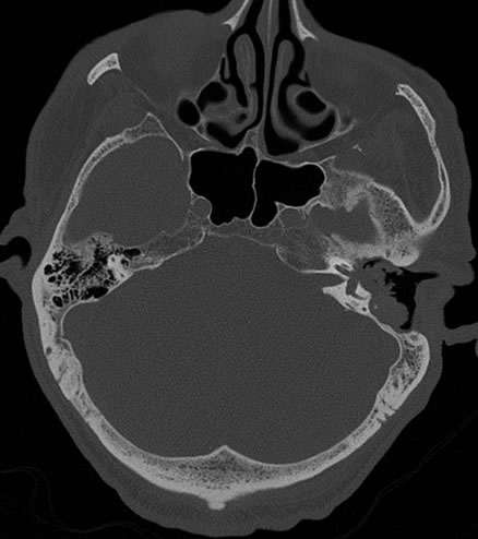
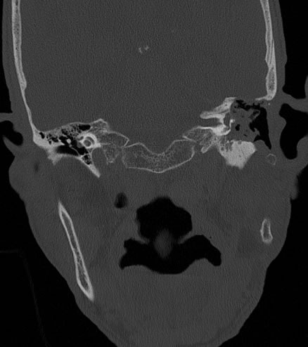
Axial and coronal CT images show a large irregular peripherally well-defined soft tissue mass in the region of the left middle ear cavity and external auditory canal. The mass has caused extensive surrounding bone destruction. There is erosion through the tegmen tympani, destruction of the otic capsule with marked erosion of the vestibule, partial erosion of the lateral semicircular canal and anterior margin of the superior semicircular canal, and thinning of the bone margin surrounding the left cochlea. The left middle ear ossicles are absent.
Differential Diagnosis:
Cholesteatoma, squamous cell carcinoma
Discussion:
Cholesteatomas are soft tissue masses composed of exfoliated keratin that is lined by stratified squamous epithelium. Cholesteatomas do not represent a malignant process. They are believed to arise when a retraction pocket of skin is formed which then becomes clogged. This most commonly occurs in the area of the pars flaccida in Prussak's space. The squamous cells that are trapped inside continue to produce keratin, which causes the mass to expand. As they grow, they may cause local bone destruction, as in the case above. The best imaging for cholesteatomas is a CT through the temporal bone, which will show a unilateral soft tissue mass that may include local bony destruction. Half of cholesteatomas will exhibit bone fragments or flecks within the mass. Cholesteatomas typically present in the fifth to eighth decades of life as otorrhea, otalgia, and conductive hearing loss. Small lesions may be treated with periodic office debridement, but larger lesions need to be surgically excised to prevent further bony destruction.
Case contributed by Christopher Heald, UC M4
BACK TO
MAIN PAGE