
Semicircular Canal Dehiscence
Findings:
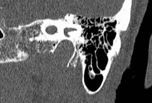
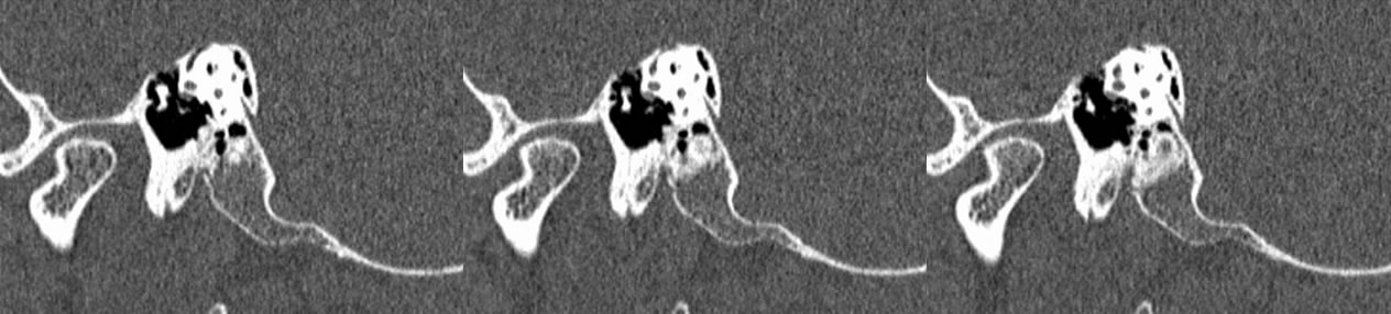
Coronal CT images demonstrate dehiscence of both superior semicircular canals. The dehiscence appears fairly wide bilaterally, slightly more evident on the right than the left.
Discussion:
Semicircular canal dehiscence describes a state in which there is extreme thinning or absence of the bony roof overlying the superior or posterior semicurcular canals. It is thought that this disease may have a congenital component that causes a thinned semicircular canal wall at birth, but has also been seen after significant head trauma. The mean age of diagnosis is 50 years old. The classic clinical findings are that of vestibular and eye movement symptoms with changes in pressure or loud sounds. An eponymous form of these symptoms is called Tullio phenomenon, which is vertigo and nystagmus that occurs with sound. These symptoms are caused by abberant movement of endolymph after compression of the oval window. Other typical signs include chronic disequilibrium, oscillopsia, and pulsatile tinnitus. The best imaging modality is CT which shows extreme thinning or absence of the roof of the superior semicircular canal or superficial bony wall of the posterior semicircular canal. Treatment of this condition in the short term involves earplugs and avoid provoking stimuli. Definitive treatment is surgical and involves resurfacing the affected semicircular canal.
Case contributed by Christopher Heald, UC M4
BACK TO
MAIN PAGE