-early cerebritis (3-5d), late cerebritis (2 wks), early capsule (>2 wks), late capsule (wks-mos)
-only 50% have fever or WBC count, 90% have headache, most have high ESR
-less than 2.5 cm may be treated with Abx alone, greater need drainage
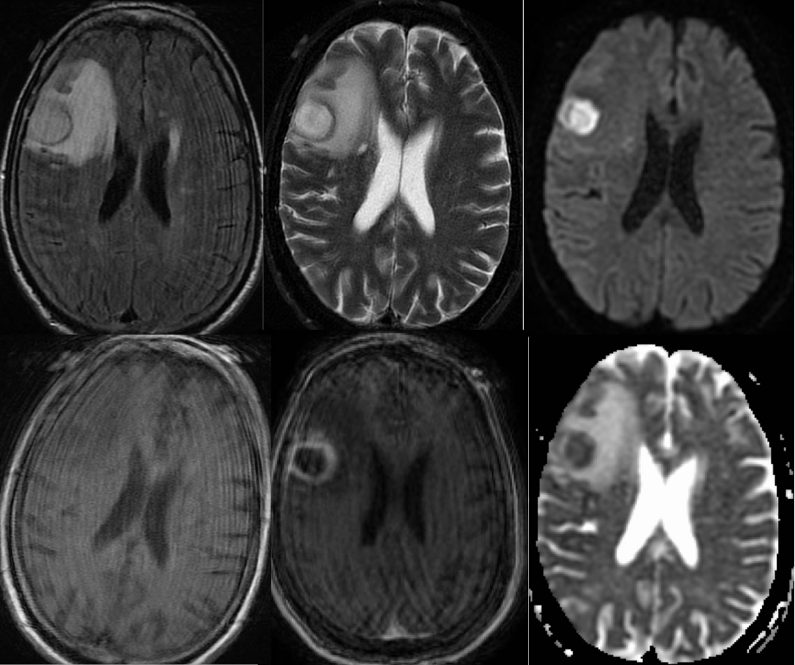
-internal DWI hyperintensity characteristic, some help in dist GBM/mets from abscess, not absolute
-DWI may be helpful in f/u, development of hyper DWI may indicate recurrence
-T2 hypointense rim with late cerebritis and early capsule, also not absolute