Gliosarcoma
Findings:
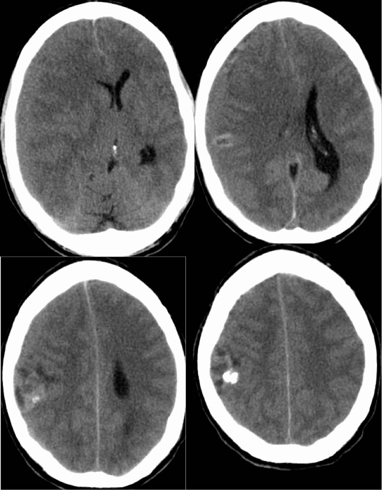
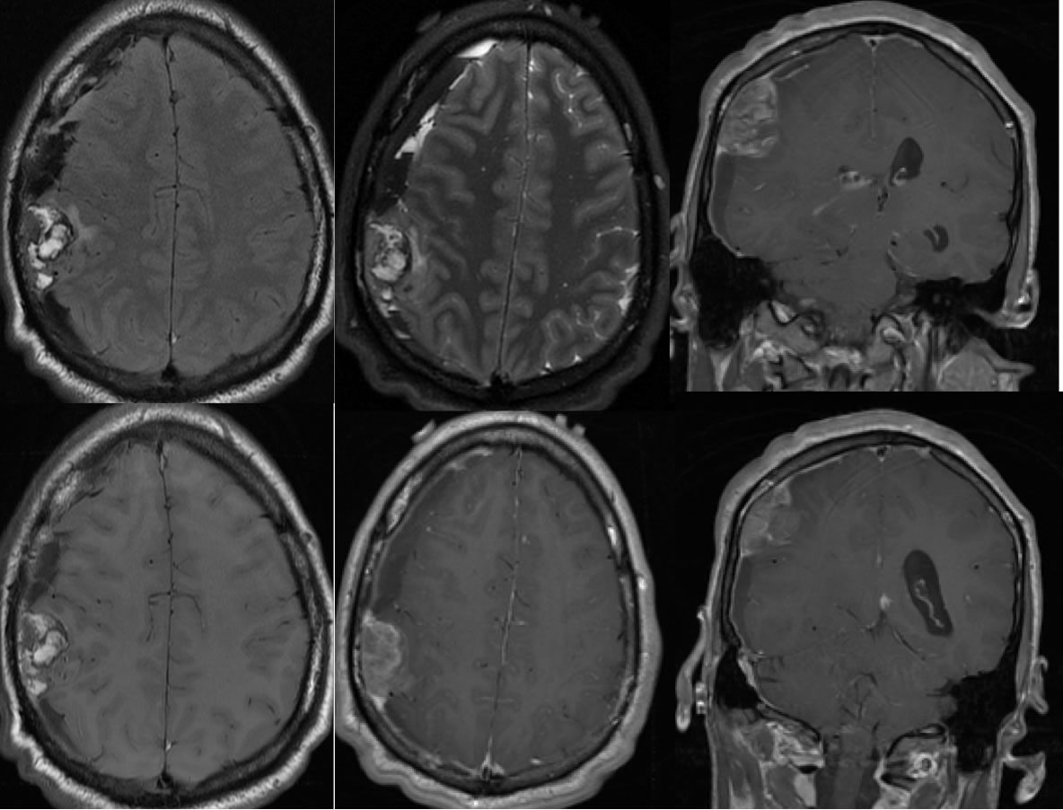
Axial noncontrast CT images demonstrate a moderate sized generally isodense subdural hematoma over the right cerebral convexity causing significant mass effect and midline shift. Also associated with the hematoma there is a heterogeneous mass along the right frontoparietal convexity with calcifications and mixed density. The MR images demonstrate a complex subdural hematoma over the right cerebral convexity. A heterogeneously enhancing mass along the right lateral frontoparietal region appears extra-axial with dural tails on either side of the lesion. The mass demonstrates marked heterogeneous signal. The medial margin of the lesion is inseparable from the cortical surface and cortical invasion or origin of the lesion could be considered.
Differential Diagnosis:
PNET, aggressive/malignant meningioma, metastasis, dural sarcoma
Discussion:
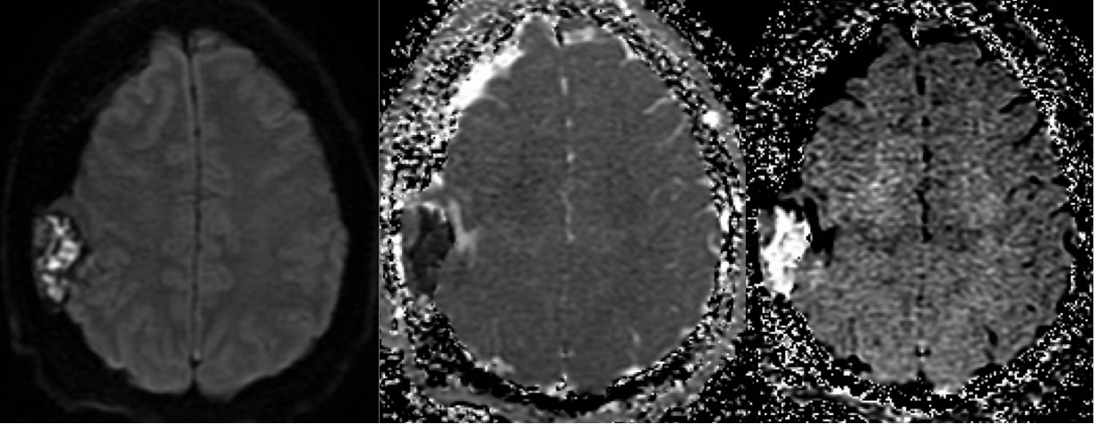
Primitive neuroectodermal tumor (PNET) is an uncommon aggressive malignancy histologically composed of "small round blue cells" that is typically found in children and young adults. Of PNETs, medulloblastoma (PNET-MB) occuring in the posterior fossa is most common. Pineoblastoma is the most common supratentorial PNET and spinal PNET is rare. In this case, the tumor is extraaxial and more appropriately considered an even more rare peripheral rather than a CNS PNET. The lesion shows a variegated histology with partial differentiation into neuroglial and potentially mesenchymal cell lines which correlates with the marked heterogenous imaging appearance. The mass may be commonly partially calcified and with cystic and solid components. Hypercellularity causes restricted diffusion. Intraaxial lesions often show less than expected edema for the size and heterogeneity of the tumor. Dissemination is common and prognosis is poorer for supratentorial/extraaxial PNET than the more common PNET-MB.
**please note that for this case the initial frozen section diagnosis was PNET, but the final pathology was read as gliosarcoma.
BACK TO
MAIN PAGE