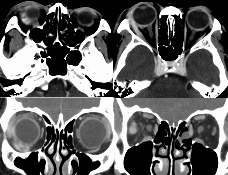
Orbital Pseudotumor (AKA Idiopathic Orbital Inflammatory Syndrome)
-idiopathic nongranulomatous inflammatory disorder
-third most common orbital disease (5%)
-children 15% of all cases
-acute onset with pain, swelling, erythema, ptosis, painful/restricted eye movement
-acute form usually responds to steroids
-uncommonly chronic with diplopia, proptosis
-chronic less commonly responds to steroids- XRT or chemo may help
-involves lacrimal gland, EOMs, fat
-orbital apex, cavernous sinus= Tolosa Hunt- painful ophthalmoplegia
-other systemic diseases associated:
-Wegener's, PAN, RP fibrosis, PSC, Reidel's, SLE, RA, dermatomyositis
Imaging:
-tendons involved and unilateral (to distinguish from Graves)
-marked enhancement
-retrobulbar fat stranding
-may present as focal or infiltrating mass
-rare bone destruction
-T2 hypointensity to distinguish from mets