
Global anoxic/hypoxic injury, subacute
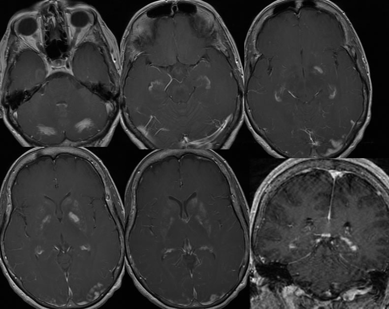
Findings:
Multiple MR images demonstrate a patchy focus of T2 hyperintensity within the left lentiform nucleus. There is no definite restricted diffusion. On the post contrast MRI images, there is nearly symmetric patchy enhancement of the bilateral hippocampal cortex, bilateral basal ganglia, caudate nuclei, and cerebellar white matter. Additional foci of gyriform enhancement involve the bilateral parietooccipital regions, left greater than right.
Differential Diagnosis:
subacute hypoxic/ischemic injury, mutiple subacute infarcts of other cause, multifocal encephalitis.
Discussion:
This patient was "found down" for unknown duration and had significantly depressed respirations and hypoxia when evaluated by the squad. At the time of MR evaluation 8 days after the insult, the patient has severe short term memory loss and visual field defects but no gross motor deficit.
Global hypoxic/ischemic injuries may have a variable appearance depending on age of the patient, type and duration of insult, and comorbidities. In this case, there is predominant involvement of deep brain structures and the hippocampi.
In the acute to early subacute phase of a severe global anoxic event, such as cardiac arrest with ineffective CPR and delayed ROSC, there may be diffuse cortical laminar necrosis and symmetric infarctions of deep brain nuclei which will have a perfectly symmetric appearance on MR, but demonstrate an accentuated grey white interface and abnormal increased T2 signal of these structures. Diffuse cortical and deep grey diffusion restriction will be seen, but might be overlooked if window leveling of the images is made inappropriately wide. These scans might appear normal to those who are inexperienced MR readers.
BACK TO
MAIN PAGE