Band Heterotopia and AVM
Findings:
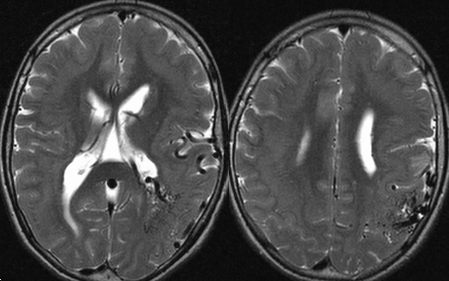
Axial T2 weighted MR images demonstrate a uniform zone of gray matter signal intensity involving the bilateral cerebral hemispheres along the subependymal zones. In addition, there is abnormal vascularity along the left temporal lobe within large feeding vessels and draining veins.
Discussion:
Regarding gray matter heterotopias, subependymal nodules are most common, but a diffuse subcortical band may also be seen. Other anomalies may be associated, such as lissencephaly or schizencephaly, including other types of cortical dysplasia. Seizures are the most common presentation along with developmental delay. The imaging appearance may have variable morphology from a minute nodule to large confluent or diffuse lesions, however these are always isointense and isodense to gray matter on all imaging. Tuberous Sclerosis may stimulate heterotopia, but other findings of TS are usually present. Tubers seen in TS may enhance and/or calcify, which is not seen with heterotopia. Etiology of these lesions is migrational arrest/slowing from subependymomal zones to the cortex. The process may be genetic as inherited or acquired mutation, or acquired in utero related to congenital infection or radiotherapy. These lesions are part of many syndromes, but small lesions may be incidental findings and asymptomatic.