Hemangiopericytoma
Findings:
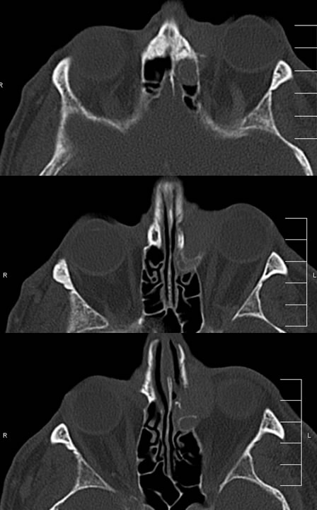
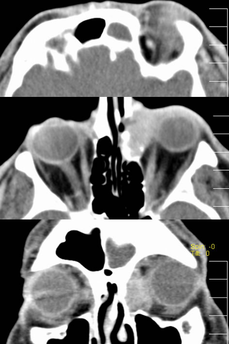
Multiple CT-bone reconstructed images demonstrate a soft tissue mass along the left medial canthus, associated with destruction of the medial orbital wall. There is some bone thickening along the posterior margin of the lesion with remodeling indicating some indolence. The lesion demonstrates diffuse enhancement after contrast administration.
Discussion:
The differential diagnosis for this process also includes a lacrimal sac adenocarcinoma, schwannoma, cavernous hemangioma, meningioma, angiosarcoma, or malignant fibrous histiocytoma. The imaging appearance is of a nonspecific aggressive mass with bone destruction and intense enhancement, less well-defined then hemangioma. Angiography may help distinguish in selected cases with an early hypervascular blush, but this is also similar to sarcomas.
The clinical features are a rare slow-growing tumor of pericytes, of which 50% are malignant, and metastases are uncommon. The lesions are treated with wide surgical excision but may recur.