CNS Lymphoma
Findings:
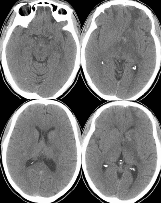
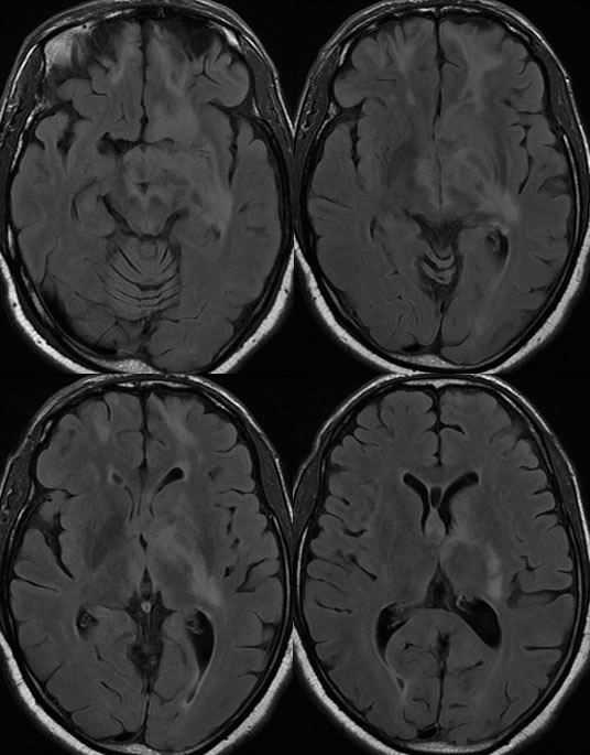
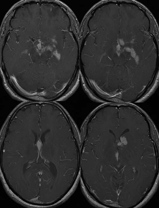
Multiple axial CT images without contrast demonstrate poorly defined low attenuation in the bilateral frontal white matter, with superimposed zones of relative hyperdensity projected over the left caudate nucleus. There is mild mass effect with partial effacement of the left frontal horn. Infiltrative low attenuation extends into the left thalamus and internal capsule as well as the lentiform nucleus. The axial FLAIR images also demonstrate the infiltrative signal abnormalities, but also confirm that the superimposed frontal signal abnormalities represent superimposed encephalomalacia related to a remote insult. After gadolinium administration, there is patchy nodular enhancement of this process which also extends into the third ventricle and suprasellar cistern.
Discussion:
Please refer to discussion of several other cases of lymphoma on this site.