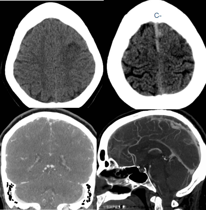
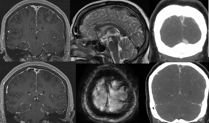
Superior sagittal sinus thrombosis with venous infarction left frontal lobe.
Findings:
Axial noncontrast CT images demonstrate increased density of the superior sagittal sinus. There is also a patchy focus of low attenuation within the right frontal lobe. On the CT venogram, there is a large elongated filling defect within the superior sagittal sinus. MR images confirm the presence of thrombosis of the superior sagittal sinus. Axial GRE images demonstrate blooming associated with the superior sagittal sinus and left frontal cortical veins. There is superimposed patchy decreased T1 signal within the left frontal lobe compatible with venous infarction.