Anaplastic Astrocytoma
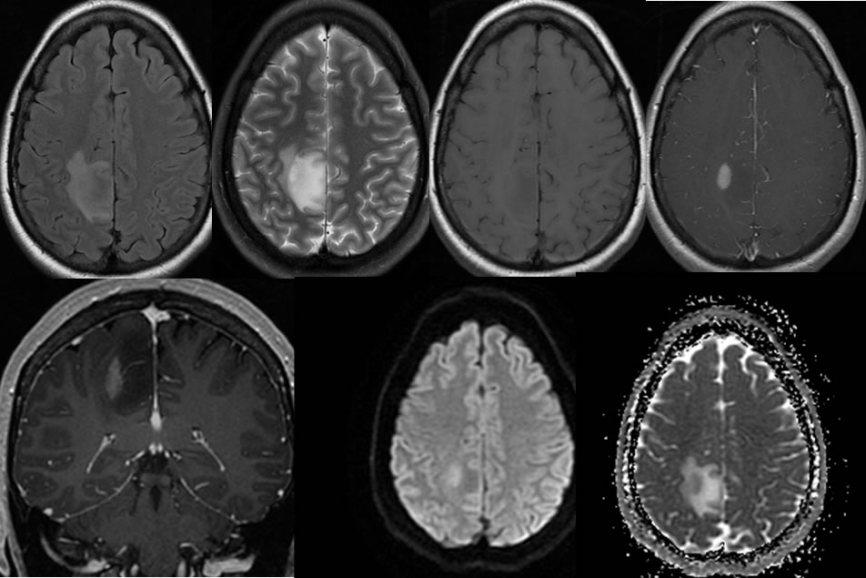
Findings:
Multiple MR images demonstrate a poorly defined signal abnormality in the right parietal parasagittal region, associated with gyral swelling. An eccentric nodular zone of enhancement is noted along the lateral aspect which demonstrates restricted diffusion.
Discussion:
-histology: well differentiated (fibrillary), anaplastic, GBM
-presence of necrosis separates GBM from anaplastic astrocytoma
-GBM may be better circumscribed microscopically than lower grade astrocytoma
-low grade (WHO I or II): children and adults 20-40, no necrosis or neovascularity, some cystic, calcification 20%, fysr 33%
-high grade (WHO III or IV): age>40, necrosis, neovascularity, hemorrhage. median sr 8 mos.
-spread- natural passages, subpial, subependymal, WM tracts, across meninges