Neurosarcoidosis
Findings:
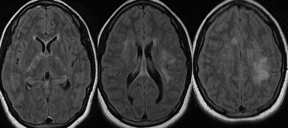
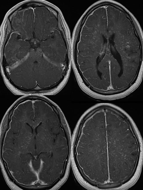
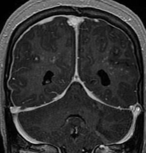
Axial FLAIR images demonstrate patchy signal abnormalities within the bilateral centrum semiovale extending into the internal capsules, slightly asymmetric in the left deep frontoparietal white matter. After gadolinium administration, there is thick dural enhancement over the right cerebral convexity. Numerous punctate enhancing lesions are present throughout the bilateral cerebral white matter which generally extend along perivascular spaces.
Discussion:
nDDx
nSarcoid, TB, mets, meningitis, (LCH)
nClinical/Path
nM>F 2:1, 3rd-4th decade; 3-5% children, 10:1 AA:CA
nEtiology unknown ?abnormal immune response or antigen
n>90% NS pts have lung disease, 5% with clinical NS (up to 27% at autopsy). Isolated NS in <1%.
n50% of CNS involvement is subclinical, may be monophasic
nNoncaseating granulomas, intraaxial or extraaxial, perivascular---->vasculitis/infarcts
nMost common complication: Hydrocephalus
nRx steroids +/- immunosuppression- but less effective for NS vs pulmonary
nImaging
nMR- diffuse or focal leptomeningeal/pachymeningeal enhancement (most common), nonspec WM lesions (MS), focal mass, basal meningeal pattern, pituitary/infundibulum, CN enhancement, hydrocephalus
BACK TO
MAIN PAGE