Global Anoxic Brain Injury
Findings:
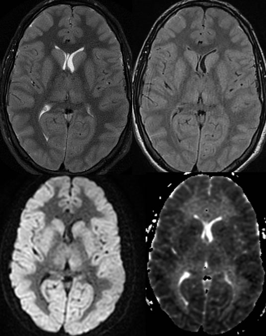
Multiple MR images demonstrate abnormal FLAIR and T2 hyperintensity of the entirety of the cerebral cortex and deep gray nuclei. These findings are also associated with diffuse restricted diffusion.
Discussion:
Clinical
n“mental status changes”- look for evidence of global insult
nSevere hypoxia at cellular level-
-Hypoperfusion, hypoxia, CO inhalation, hypotension, anoxia, CP arrest
Imaging
nLoss of GW differentiation on CT- but GWD accentuated on MR
nIncreased T2 signal of cortex and basal ganglia/thalami
nDiffuse diffusion restriction of gray matter and gray nuclei
nCareful with window leveling
nADC map- still see accentuated GWD, not seen with normal
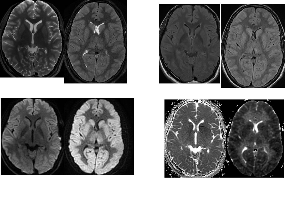
Below is a comparison of normal (L) and anoxic injury (R) MR imaging in similar age patients:
BACK TO
MAIN PAGE