Diffuse intraaxial metastases, occult on noncontrast CT
Findings:
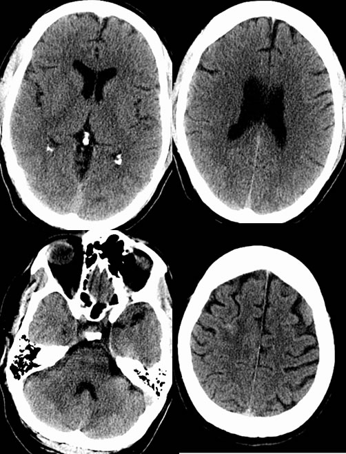
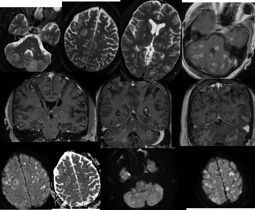
Axial noncontrast CT images demonstrate a thin linear hyperdensity in the right frontal lobe. There is slight heterogeneity of the cerebellum on the CT. The MR images performed immediately after CT demonstrate diffuse intraaxial ring enhancing lesions with no significant associated mass effect.Small zones of vasogenic edema are associated with some of the lesions.The lesions in general exhibit peripheral restricted diffusion.
Discussion:
This case illustrates the limitations of noncontrast CT for detection of significant intracranial pathology.